Single Molecule Imaging of Genome Duplication and Maintenance Laboratory, The Francis Crick Institute, NW1 1AT London, UK.
Novo Nordisk Foundation Center for Protein Research, Faculty of Health and Medical Sciences, University of Copenhagen, DK-2200 Copenhagen, Denmark.
Cell Rep. 2019 Feb 19;26(8):2113-2125.e6. doi: 10.1016/j.celrep.2019.01.086.
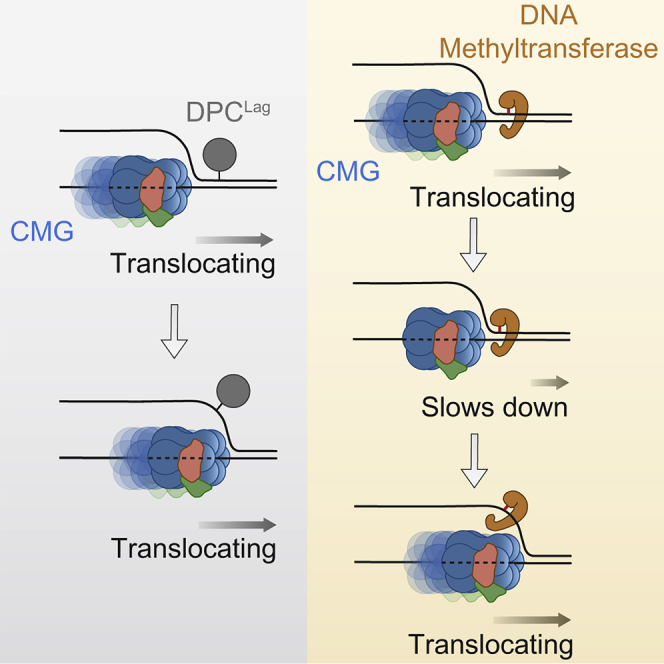
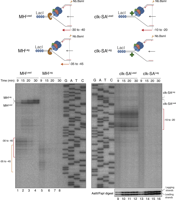
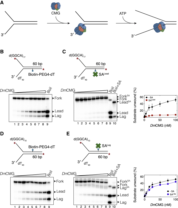
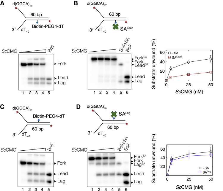
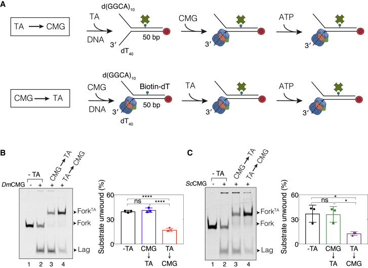
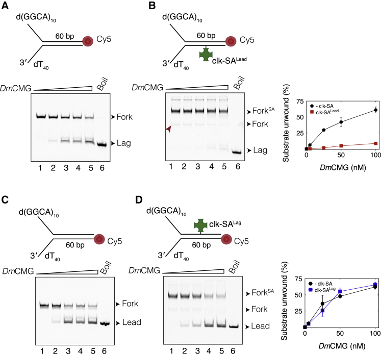
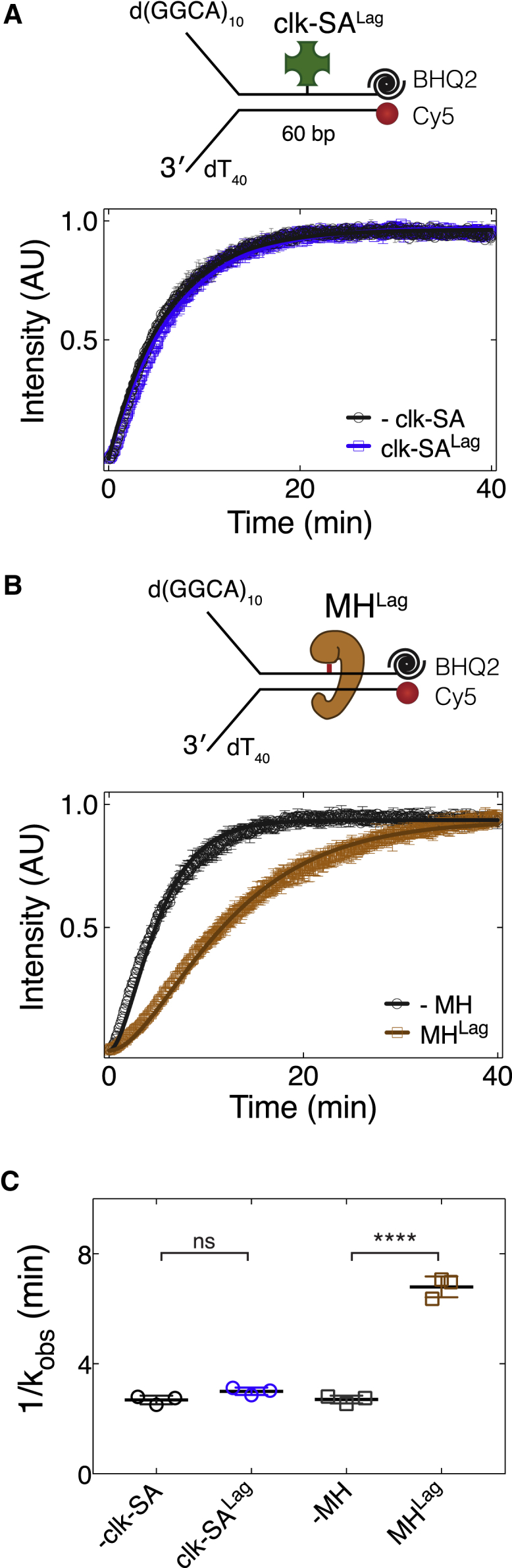
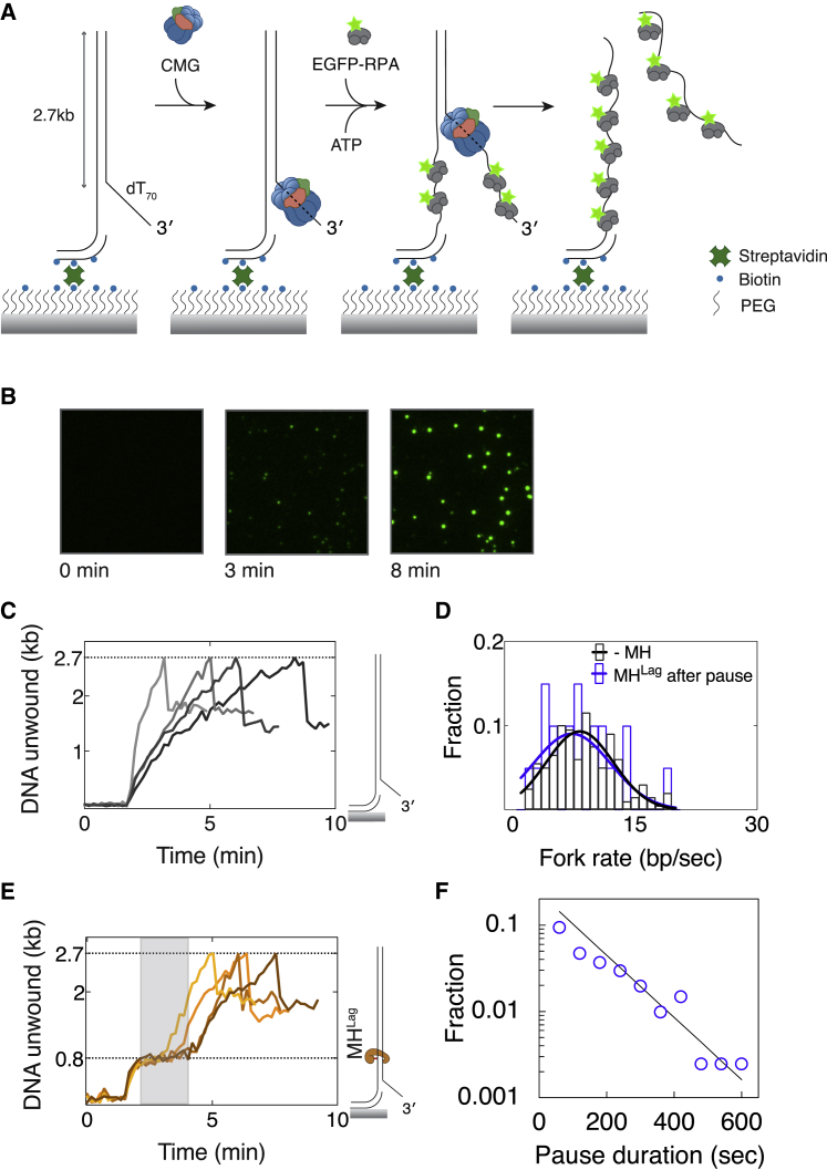
Progression of DNA replication depends on the ability of the replisome complex to overcome nucleoprotein barriers. During eukaryotic replication, the CMG helicase translocates along the leading-strand template and unwinds the DNA double helix. While proteins bound to the leading-strand template efficiently block the helicase, the impact of lagging-strand protein obstacles on helicase translocation and replisome progression remains controversial. Here, we show that CMG and replisome progressions are impaired when proteins crosslinked to the lagging-strand template enhance the stability of duplex DNA. In contrast, proteins that exclusively interact with the lagging-strand template influence neither the translocation of isolated CMG nor replisome progression in Xenopus egg extracts. Our data imply that CMG completely excludes the lagging-strand template from the helicase central channel while unwinding DNA at the replication fork, which clarifies how two CMG helicases could freely cross one another during replication initiation and termination.
DNA 复制的进展取决于复制体复合物克服核蛋白障碍的能力。在真核复制过程中,CMG 解旋酶沿着前导链模板移动并解开 DNA 双螺旋。虽然与前导链模板结合的蛋白质可以有效地阻止解旋酶,但滞后链蛋白障碍物对解旋酶迁移和复制体进展的影响仍然存在争议。在这里,我们表明,当交联到滞后链模板的蛋白质增强双链 DNA 的稳定性时,CMG 和复制体的进展会受到损害。相比之下,仅与滞后链模板相互作用的蛋白质既不会影响分离的 CMG 的迁移,也不会影响爪蟾卵提取物中的复制体进展。我们的数据表明,CMG 在复制叉处解开 DNA 的同时,将滞后链模板完全排除在解旋酶的中央通道之外,这阐明了在复制起始和终止过程中,两个 CMG 解旋酶如何能够自由地相互交叉。