Kurose Nozomu, Mizuguchi Seiya, Ohkanemasa Yoshiiku, Yamashita Manabu, Nakano Mariko, Guo Xin, Aikawa Akane, Nakada Satoko, Sasagawa Toshiyuki, Yamada Sohsuke
Department of Pathology and Laboratory Medicine, Kanazawa Medical University, Uchinada, Japan.
Department of Pathology, Kanazawa Medical University, Uchinada, Japan.
SAGE Open Med Case Rep. 2019 Feb 13;7:2050313X19828235. doi: 10.1177/2050313X19828235. eCollection 2019.
Tumor-associated tissue eosinophilia is defined as an inflammatory response with the marked infiltration of eosinophils within tumor tissues. Tumor-associated tissue eosinophilia has been reported in various organs; however, no studies have examined the detailed cytopathological findings of tumor-associated tissue eosinophilia.
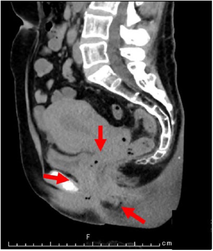
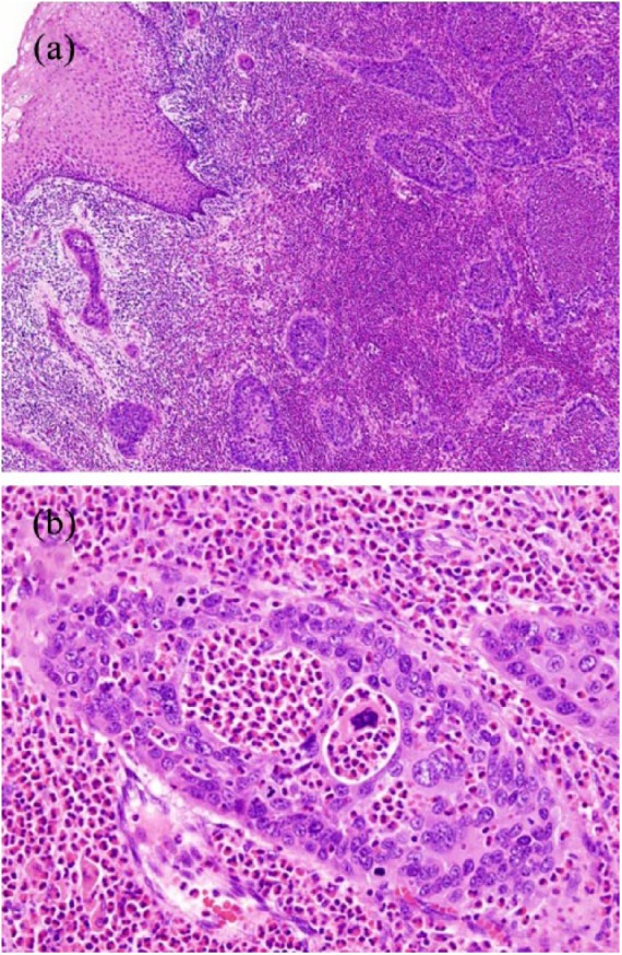
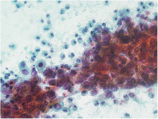
A 49-year-old woman presented with lower abdominal and back pain that had started 1 month earlier. A cervical biopsy revealed a diagnosis of non-keratinizing squamous cell carcinoma. A mildly increased number of eosinophils was observed in both cervical cytology and a biopsy. On pelvic computed tomography, a tumor mass measuring up to 5.5 cm in the largest diameter was seen in the uterine cervix. After 1 month, endometrial cytology was performed, and non-keratinizing squamous cell carcinoma together with normal endometrial glands was obtained in a background of marked eosinophil numbers. Tumor cells in an irregular-shaped solid nest had variable-sized hyperchromatic nuclei and light-green-stained cytoplasm. The number of eosinophils was obviously increased. Considering the possibility of tumor-associated tissue eosinophilia, we evaluated a peripheral blood sample and confirmed an increased number of eosinophils. Radical hysterectomy was performed, and the final pathological diagnosis was adenosquamous carcinoma. Although the number of eosinophils decreased after surgery, it increased again at the time of recurrence 1 year later. Chemo-irradiation was performed, but the patient died 1 year and 8 months after the operation.
Cytopathologists should consider the presence of tumor-associated tissue eosinophilia by focusing on not only tumor cells but also the markedly eosinophilic background. The eosinophil count might be a useful marker of the disease activity.
肿瘤相关组织嗜酸性粒细胞增多症被定义为肿瘤组织内有明显嗜酸性粒细胞浸润的炎症反应。肿瘤相关组织嗜酸性粒细胞增多症已在多个器官中被报道;然而,尚无研究对肿瘤相关组织嗜酸性粒细胞增多症的详细细胞病理学表现进行研究。
一名49岁女性,1个月前开始出现下腹部和背部疼痛。宫颈活检显示诊断为非角化性鳞状细胞癌。宫颈细胞学检查和活检均观察到嗜酸性粒细胞数量轻度增加。盆腔计算机断层扫描显示,子宫颈有一个最大直径达5.5厘米的肿瘤肿块。1个月后进行了子宫内膜细胞学检查,在嗜酸性粒细胞数量明显增多的背景下,获得了非角化性鳞状细胞癌及正常子宫内膜腺体。不规则形实性巢状结构中的肿瘤细胞有大小不一的深染核和浅绿色染色的细胞质。嗜酸性粒细胞数量明显增加。考虑到肿瘤相关组织嗜酸性粒细胞增多症的可能性,我们对一份外周血样本进行了评估,证实嗜酸性粒细胞数量增加。实施了根治性子宫切除术,最终病理诊断为腺鳞癌。虽然术后嗜酸性粒细胞数量减少,但1年后复发时又再次增加。进行了化疗放疗,但患者在手术后1年零8个月死亡。
细胞病理学家不仅应关注肿瘤细胞,还应关注明显嗜酸性粒细胞增多的背景,以考虑肿瘤相关组织嗜酸性粒细胞增多症的存在。嗜酸性粒细胞计数可能是疾病活动的一个有用标志物。