Department of Pharmacology and Experimental Neuroscience, University of Nebraska Medical Center, 985880 Nebraska Medical Center, Omaha, NE, 68198-5880, USA.
Central Institute for Experimental Animals, Kawasaki-ku, Kawasaki, Japan.
Mol Neurodegener. 2019 Mar 5;14(1):12. doi: 10.1186/s13024-019-0311-y.
Microglia are the principal innate immune defense cells of the centeral nervous system (CNS) and the target of the human immunodeficiency virus type one (HIV-1). A complete understanding of human microglial biology and function requires the cell's presence in a brain microenvironment. Lack of relevant animal models thus far has also precluded studies of HIV-1 infection. Productive viral infection in brain occurs only in human myeloid linage microglia and perivascular macrophages and requires cells present throughout the brain. Once infected, however, microglia become immune competent serving as sources of cellular neurotoxic factors leading to disrupted brain homeostasis and neurodegeneration.
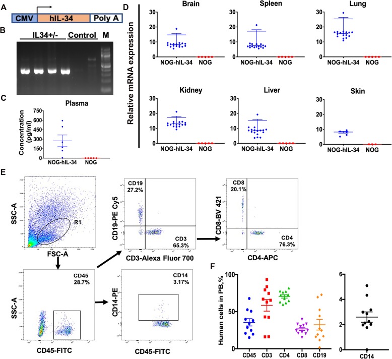
Herein, we created a humanized bone-marrow chimera producing human "microglia like" cells in NOD.Cg-PrkdcIl2rgTg(CMV-IL34)1/Jic mice. Newborn mice were engrafted intrahepatically with umbilical cord blood derived CD34+ hematopoietic stem progenitor cells (HSPC). After 3 months of stable engraftment, animals were infected with HIV-1, a myeloid-specific tropic viral isolate. Virologic, immune and brain immunohistology were performed on blood, peripheral lymphoid tissues, and brain.
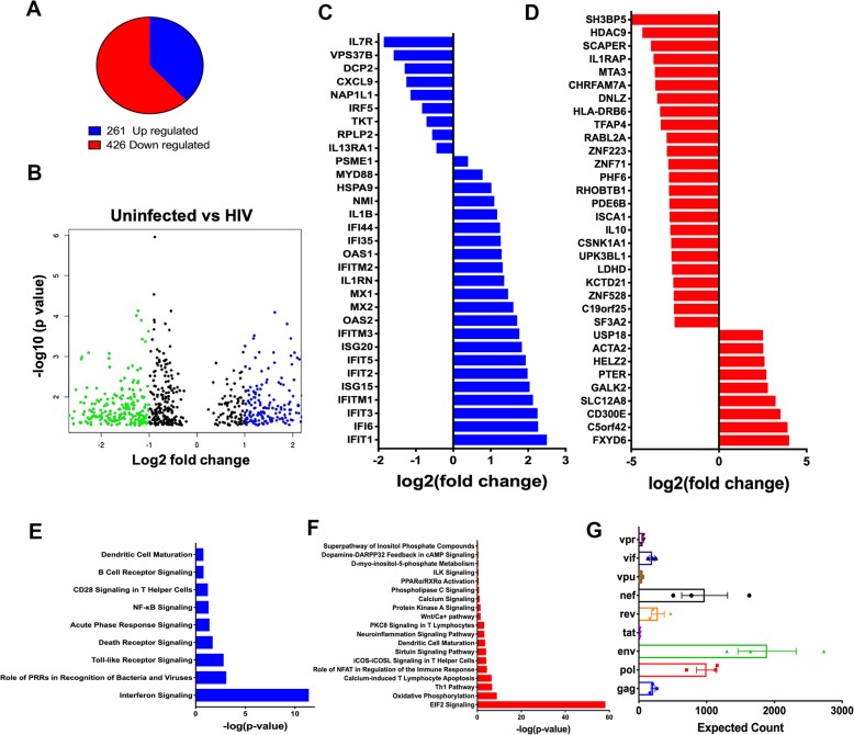
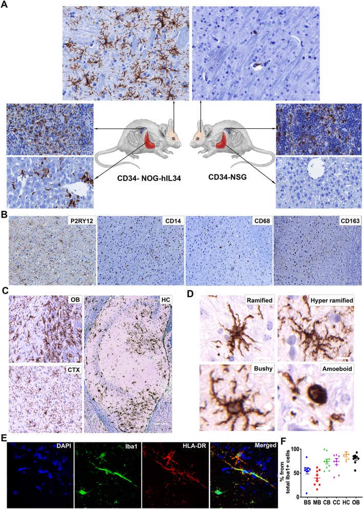
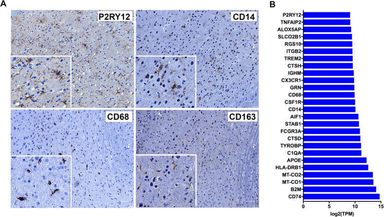
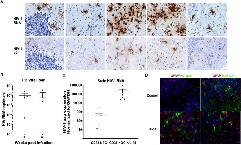
Human interleukin-34 under the control of the cytomegalovirus promoter inserted in NSG mouse strain drove brain reconstitution of HSPC derived peripheral macrophages into microglial-like cells. These human cells expressed canonical human microglial cell markers that included CD14, CD68, CD163, CD11b, ITGB2, CX3CR1, CSFR1, TREM2 and P2RY12. Prior restriction to HIV-1 infection in the rodent brain rested on an inability to reconstitute human microglia. Thus, the natural emergence of these cells from ingressed peripheral macrophages to the brain could allow, for the first time, the study of a CNS viral reservoir. To this end we monitored HIV-1 infection in a rodent brain. Viral RNA and HIV-1p24 antigens were readily observed in infected brain tissues. Deep RNA sequencing of these infected mice and differential expression analysis revealed human-specific molecular signatures representative of antiviral and neuroinflammatory responses.
This humanized microglia mouse reflected human HIV-1 infection in its known principal reservoir and showed the development of disease-specific innate immune inflammatory and neurotoxic responses mirroring what can occur in an infected human brain.
小胶质细胞是中枢神经系统(CNS)中主要的先天免疫防御细胞,也是人类免疫缺陷病毒 1 型(HIV-1)的靶标。要完全了解人类小胶质细胞的生物学和功能,需要将细胞置于脑微环境中。迄今为止,缺乏相关的动物模型也妨碍了对 HIV-1 感染的研究。只有在人脑髓系谱系小胶质细胞和血管周巨噬细胞中才会发生病毒的有效感染,并且需要存在于整个大脑中的细胞。然而,一旦被感染,小胶质细胞就会变得具有免疫能力,成为导致脑内稳态破坏和神经退行性变的细胞神经毒性因子的来源。
在此,我们创建了一种人源化骨髓嵌合体,在 NOD.Cg-PrkdcIl2rgTg(CMV-IL34)1/Jic 小鼠中产生具有“小胶质细胞样”的细胞。新生小鼠经肝内移植脐带血衍生的 CD34+造血干细胞祖细胞(HSPC)。稳定植入 3 个月后,动物感染 HIV-1,这是一种髓系特异性的病毒分离株。对血液、外周淋巴组织和大脑进行病毒学、免疫和脑免疫组织化学检测。
受巨细胞病毒启动子控制的人白细胞介素 34 插入 NSG 小鼠品系中,驱动 HSPC 衍生的外周巨噬细胞向小胶质细胞样细胞在大脑中的再构成。这些人类细胞表达了经典的人类小胶质细胞标志物,包括 CD14、CD68、CD163、CD11b、ITGB2、CX3CR1、CSFR1、TREM2 和 P2RY12。以前,由于不能重建人类小胶质细胞,限制了 HIV-1 在啮齿动物大脑中的感染。因此,从入侵的外周巨噬细胞到大脑中自然出现这些细胞,可以首次研究中枢神经系统病毒库。为此,我们监测了啮齿动物大脑中的 HIV-1 感染。在受感染的脑组织中很容易观察到病毒 RNA 和 HIV-1p24 抗原。对这些感染小鼠进行深度 RNA 测序和差异表达分析,揭示了代表抗病毒和神经炎症反应的人类特异性分子特征。
这种人源化小胶质细胞小鼠反映了其主要病毒库中的人类 HIV-1 感染,并表现出与感染人类大脑中发生的情况相类似的特定疾病的先天免疫炎症和神经毒性反应。