Oladnabi Morteza, Bagheri Abouzar, Rezaei Kanavi Mozhgan, Azadmehr Abbas, Kianmehr Anvarsadat
Stem Cell Research Center, Golestan University of Medical Sciences, Gorgan, Iran.
Ischemic Disorders Research Center, Golestan University of Medical Sciences, Gorgan, Iran.
Iran J Basic Med Sci. 2019 Feb;22(2):128-133. doi: 10.22038/ijbms.2018.25023.6214.
It is known that extremely low frequency-pulsed electromagnetic fields (ELF-PEMF) influence multiple cellular and molecular processes. Retinal pigment epithelial (RPE) cells have a significant part in the emergence and pathophysiology of several ocular disorders, such as neovascularization. This study assessed the impact of ELF-PEMF on the proangiogenic features of RPE cells.
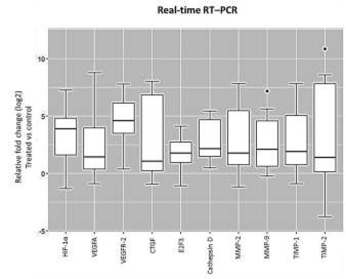
Primary cultured RPE cells were treated with ELF-PEMF (50 Hz) for three days. Using ELISA assay, we evaluated the effects of treatment on RPE cell proliferation and apoptosis. Also, RT-PCR was used to determine the gene expression of proangiogenic factors, such as matrix metalloproteinase-2 (MMP-2), MMP-9, vascular endothelial growth factors receptor 2 (VEGFR-2), hypoxia-inducible factor 1 (HIF-1α), VEGFA, cathepsin D, connective tissue growth factor (CTGF), E2F3, tissue inhibitors of metalloproteinases 1 (TIMP-1), and TIMP-2.
No noticeable changes were observed in cell proliferation and cell death of ELF-PEMF-exposed RPE cells, while transcript levels of proangiogenic genes (HIF-1α, VEGFA, VEGFR-2, CTGF, cathepsin D, TIMP-1, E2F3, MMP-2, and MMP-9) increased significantly.
RPE cells are important for homeostasis of the retina. ELF-PEMF increased the gene expression of proangiogenic factors in RPE cells, which highlights concerns about the impact of this treatment on human health.
已知极低频脉冲电磁场(ELF-PEMF)会影响多种细胞和分子过程。视网膜色素上皮(RPE)细胞在多种眼部疾病(如新生血管形成)的发生和病理生理过程中起重要作用。本研究评估了ELF-PEMF对RPE细胞促血管生成特性的影响。
原代培养的RPE细胞用ELF-PEMF(50Hz)处理三天。使用酶联免疫吸附测定(ELISA)法,我们评估了处理对RPE细胞增殖和凋亡的影响。此外,逆转录聚合酶链反应(RT-PCR)用于测定促血管生成因子的基因表达,如基质金属蛋白酶-2(MMP-2)、MMP-9、血管内皮生长因子受体2(VEGFR-2)、缺氧诱导因子1(HIF-1α)、VEGFA、组织蛋白酶D、结缔组织生长因子(CTGF)、E2F3、金属蛋白酶组织抑制剂1(TIMP-1)和TIMP-2。
暴露于ELF-PEMF的RPE细胞的细胞增殖和细胞死亡未观察到明显变化,而促血管生成基因(HIF-1α、VEGFA、VEGFR-2、CTGF、组织蛋白酶D、TIMP-1、E2F3、MMP-2和MMP-9)的转录水平显著增加。
RPE细胞对视网膜的稳态很重要。ELF-PEMF增加了RPE细胞中促血管生成因子的基因表达,这凸显了对这种治疗对人类健康影响的担忧。