Liu Rongrong, Winkelmann James A, Spicer Graham, Zhu Yunxiao, Eid Aya, Ameer Guillermo A, Backman Vadim, Yi Ji
1Department of Biomedical Engineering, Northwestern University, Evanston, IL 60208 USA.
2Department of Chemical and Biological Engineering, Northwestern University, Evanston, IL 60208 USA.
Light Sci Appl. 2018 Aug 29;7:57. doi: 10.1038/s41377-018-0057-2. eCollection 2018.
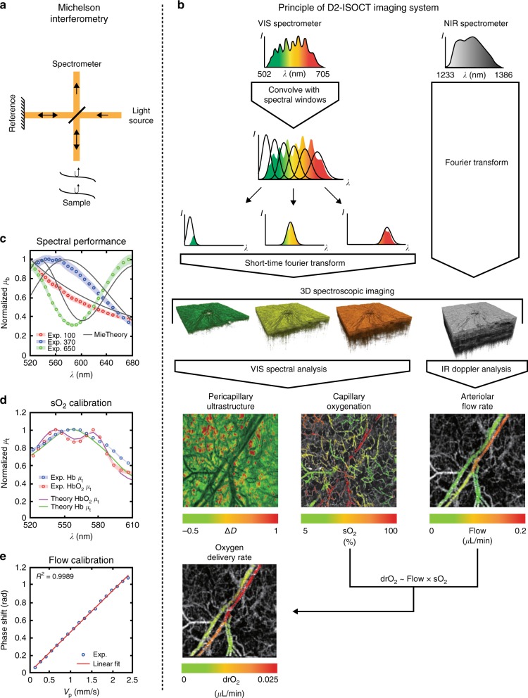
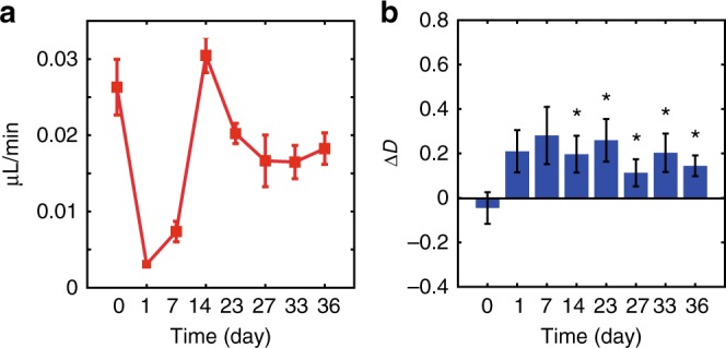
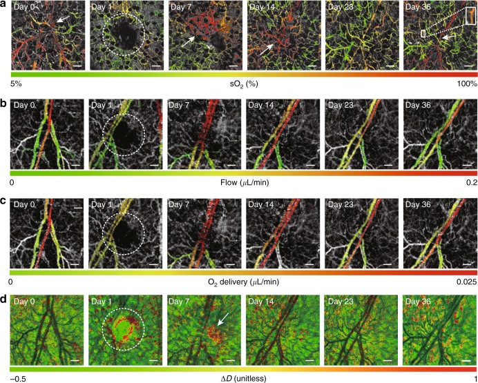
Measuring capillary oxygenation and the surrounding ultrastructure can allow one to monitor a microvascular niche and better understand crucial biological mechanisms. However, capillary oximetry and pericapillary ultrastructure are challenging to measure in vivo. Here we demonstrate a novel optical imaging system, dual-band dual-scan inverse spectroscopic optical coherence tomography (D2-ISOCT), that, for the first time, can simultaneously obtain the following metrics in vivo using endogenous contrast: (1) capillary-level oxygen saturation and arteriolar-level blood flow rates, oxygen delivery rates, and oxygen metabolic rates; (2) spatial characteristics of tissue structures at length scales down to 30 nm; and (3) morphological images up to 2 mm in depth. To illustrate the capabilities of D2-ISOCT, we monitored alterations to capillaries and the surrounding pericapillary tissue (tissue between the capillaries) in the healing response of a mouse ear wound model. The obtained microvascular and ultrastructural metrics corroborated well with each other, showing the promise of D2-ISOCT for becoming a powerful new non-invasive imaging tool.
测量毛细血管氧合作用及其周围的超微结构能够让人监测微血管生态位,并更好地理解关键的生物学机制。然而,在体内测量毛细血管血氧饱和度和毛细血管周围超微结构具有挑战性。在此,我们展示了一种新型光学成像系统,即双波段双扫描反向光谱光学相干断层扫描技术(D2-ISOCT),它首次能够利用内源性对比剂在体内同时获取以下指标:(1)毛细血管水平的氧饱和度以及小动脉水平的血流速度、氧输送速率和氧代谢速率;(2)低至30纳米长度尺度的组织结构的空间特征;(3)深度达2毫米的形态学图像。为了说明D2-ISOCT的功能,我们在小鼠耳部伤口模型的愈合反应中监测了毛细血管及其周围毛细血管周围组织(毛细血管之间的组织)的变化。所获得的微血管和超微结构指标相互印证良好,显示出D2-ISOCT有望成为一种强大的新型非侵入性成像工具。