Department of Medicine, Division of Pulmonary and Critical Care Medicine, Massachusetts General Hospital, Harvard Medical School, Boston, MA, USA.
Center for Immunology and Inflammatory Diseases, Division of Rheumatology, Allergy and Immunology, Massachusetts General Hospital, Harvard Medical School, Boston, MA, USA.
Respirology. 2019 Nov;24(11):1073-1080. doi: 10.1111/resp.13521. Epub 2019 Mar 7.
In vivo evaluation of the microstructural differences between asthmatic and non-asthmatic airways and their functional consequences is relevant to understanding and, potentially, treating asthma. In this study, we use endobronchial optical coherence tomography to investigate how allergic airways with asthma differ from allergic non-asthmatic airways in baseline microstructure and in response to allergen challenge.
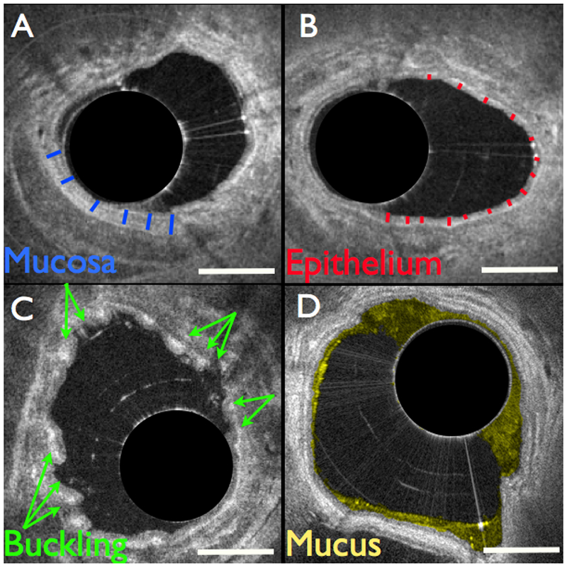
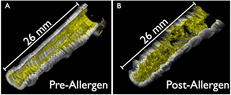
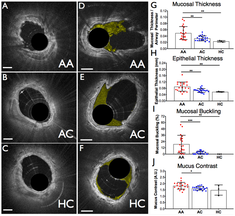
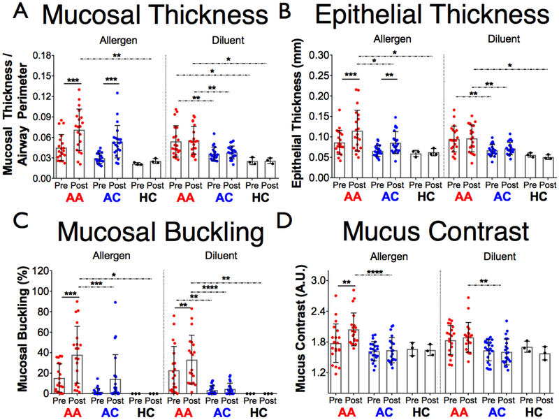
A total of 45 subjects completed the study, including 20 allergic, mildly asthmatic individuals, 22 non-asthmatic allergic controls and 3 healthy controls. A 3-cm airway segment in the right middle and right upper lobe were imaged in each subject immediately before and 24 h following segmental allergen challenge to the right middle lobe. Relationships between optical airway measurements (epithelial and mucosal thicknesses, mucosal buckling and mucus) and airway obstruction (FEV /FVC (forced expiratory volume in 1 s/forced vital capacity) and FEV % (FEV as a percentage of predictive value)) were investigated.
Significant increases at baseline and in response to allergen were observed for all four of our imaging metrics in the asthmatic airways compared to the non-asthmatic airways. Epithelial thickness and mucosal buckling exhibited a significant relationship to FEV /FVC in the asthmatic group.
Simultaneous assessments of airway microstructure, buckling and mucus revealed both structural and functional differences between the mildly asthmatic and control groups, with airway buckling seeming to be the most relevant factor. The results of this study demonstrate that a comprehensive, microstructural approach to assessing the airways may be important in future asthma studies as well as in the monitoring and treatment of asthma.
对哮喘和非哮喘气道之间的微观结构差异及其功能后果进行体内评估,与理解(并潜在地治疗)哮喘有关。在这项研究中,我们使用支气管内光学相干断层扫描来研究具有哮喘的过敏性气道与具有过敏但非哮喘的气道在基线微观结构以及对变应原挑战的反应方面有何不同。
共有 45 名受试者完成了该研究,包括 20 名过敏性轻度哮喘患者、22 名非哮喘过敏性对照者和 3 名健康对照者。在右中叶和右上叶的每个气道段进行 3cm 的成像,在右中叶节段变应原挑战前和 24 小时后立即对每个受试者进行成像。研究了光学气道测量值(上皮和粘膜厚度、粘膜弯曲和粘液)与气道阻塞(FEV/FVC(1 秒用力呼气量/用力肺活量)和 FEV%(FEV 作为预测值的百分比))之间的关系。
与非哮喘气道相比,哮喘气道的所有四个成像指标在基线和对变应原的反应中均有显著增加。上皮厚度和粘膜弯曲与哮喘组的 FEV/FVC 呈显著相关。
同时评估气道的微观结构、弯曲和粘液显示了轻度哮喘组和对照组之间的结构和功能差异,气道弯曲似乎是最相关的因素。这项研究的结果表明,全面的、微观结构的气道评估方法可能在未来的哮喘研究以及哮喘的监测和治疗中很重要。