• 文献检索
  • 文档翻译
  • 深度研究
  • 学术资讯
  • Suppr Zotero 插件Zotero 插件
  • 邀请有礼
  • 套餐&价格
  • 历史记录
应用&插件
Suppr Zotero 插件Zotero 插件浏览器插件Mac 客户端Windows 客户端微信小程序
定价
高级版会员购买积分包购买API积分包
服务
文献检索文档翻译深度研究API 文档MCP 服务
关于我们
关于 Suppr公司介绍联系我们用户协议隐私条款
关注我们

Suppr 超能文献

核心技术专利:CN118964589B侵权必究
粤ICP备2023148730 号-1Suppr @ 2026

文献检索

告别复杂PubMed语法,用中文像聊天一样搜索,搜遍4000万医学文献。AI智能推荐,让科研检索更轻松。

立即免费搜索

文件翻译

保留排版,准确专业,支持PDF/Word/PPT等文件格式,支持 12+语言互译。

免费翻译文档

深度研究

AI帮你快速写综述,25分钟生成高质量综述,智能提取关键信息,辅助科研写作。

立即免费体验

格里森概率图谱:一种用于在MRI空间中绘制前列腺癌可能性的放射组学工具。

Gleason Probability Maps: A Radiomics Tool for Mapping Prostate Cancer Likelihood in MRI Space.

作者信息

McGarry Sean D, Bukowy John D, Iczkowski Kenneth A, Unteriner Jackson G, Duvnjak Petar, Lowman Allison K, Jacobsohn Kenneth, Hohenwalter Mark, Griffin Michael O, Barrington Alex W, Foss Halle E, Keuter Tucker, Hurrell Sarah L, See William A, Nevalainen Marja T, Banerjee Anjishnu, LaViolette Peter S

机构信息

Departments of Radiology.

Pathology.

出版信息

Tomography. 2019 Mar;5(1):127-134. doi: 10.18383/j.tom.2018.00033.

DOI:10.18383/j.tom.2018.00033
PMID:30854450
原文链接:https://pmc.ncbi.nlm.nih.gov/articles/PMC6403022/
Abstract

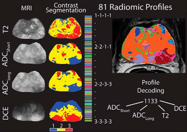
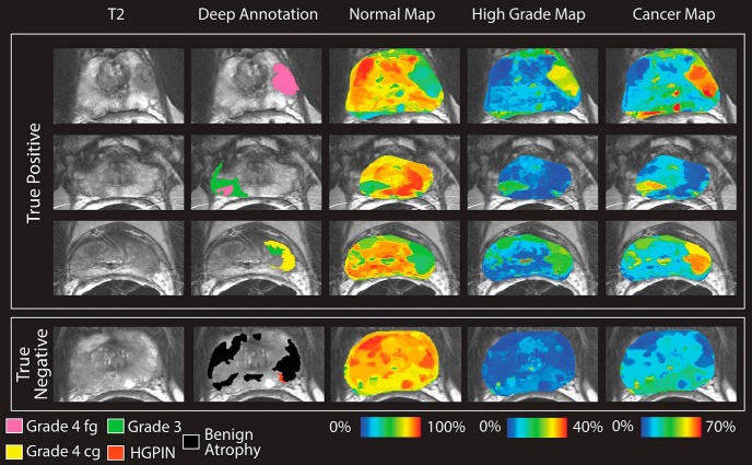
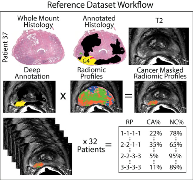
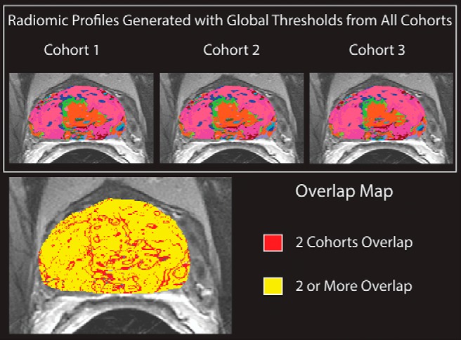
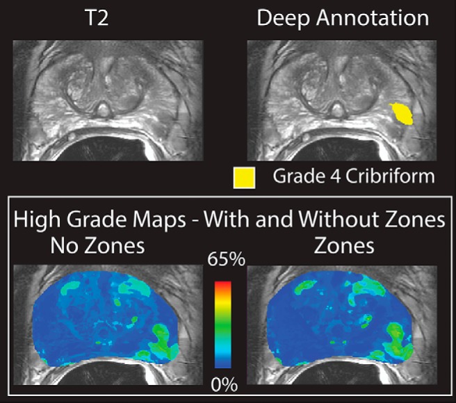
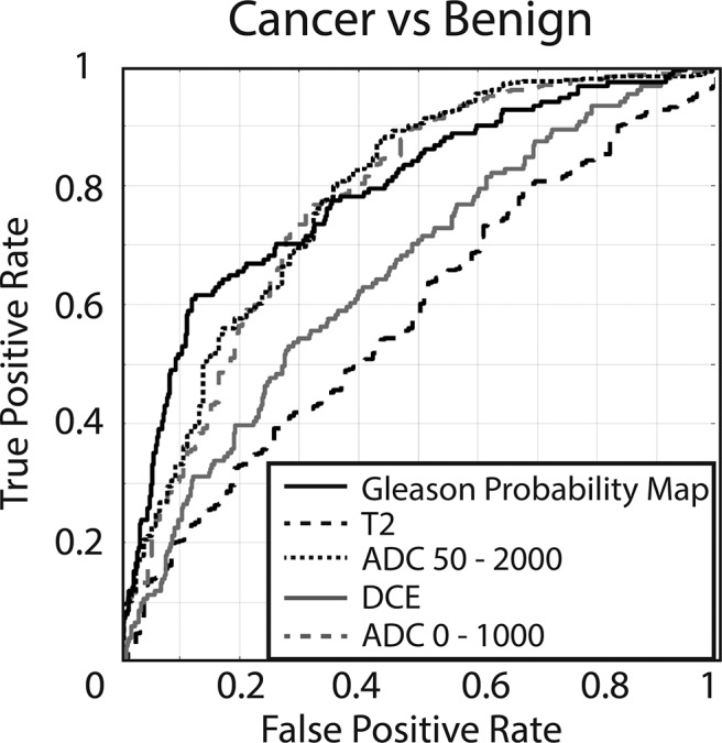
Prostate cancer is the most common noncutaneous cancer in men in the United States. The current paradigm for screening and diagnosis is imperfect, with relatively low specificity, high cost, and high morbidity. This study aims to generate new image contrasts by learning a distribution of unique image signatures associated with prostate cancer. In total, 48 patients were prospectively recruited for this institutional review board-approved study. Patients underwent multiparametric magnetic resonance imaging 2 weeks before surgery. Postsurgical tissues were annotated by a pathologist and aligned to the in vivo imaging. Radiomic profiles were generated by linearly combining 4 image contrasts (T2, apparent diffusion coefficient [ADC] 0-1000, ADC 50-2000, and dynamic contrast-enhanced) segmented using global thresholds. The distribution of radiomic profiles in high-grade cancer, low-grade cancer, and normal tissues was recorded, and the generated probability values were applied to a naive test set. The resulting Gleason probability maps were stable regardless of training cohort, functioned independent of prostate zone, and outperformed conventional clinical imaging (area under the curve [AUC] = 0.79). Extensive overlap was seen in the most common image signatures associated with high- and low-grade cancer, indicating that low- and high-grade tumors present similarly on conventional imaging.

摘要

前列腺癌是美国男性中最常见的非皮肤癌。目前的筛查和诊断模式并不完美,具有相对较低的特异性、高成本和高发病率。本研究旨在通过学习与前列腺癌相关的独特图像特征分布来生成新的图像对比。总共前瞻性招募了48名患者参与这项经机构审查委员会批准的研究。患者在手术前2周接受多参数磁共振成像检查。术后组织由病理学家进行标注,并与活体成像进行比对。通过线性组合使用全局阈值分割的4种图像对比(T2、表观扩散系数[ADC] 0 - 1000、ADC 50 - 2000和动态对比增强)生成放射组学特征。记录高级别癌、低级别癌和正常组织中放射组学特征的分布,并将生成的概率值应用于一个朴素测试集。无论训练队列如何,所得的 Gleason 概率图都很稳定,与前列腺区域无关,并且优于传统临床成像(曲线下面积[AUC] = 0.79)。在与高级别癌和低级别癌相关的最常见图像特征中发现了广泛的重叠,这表明低级别和高级别肿瘤在传统成像上表现相似。

https://cdn.ncbi.nlm.nih.gov/pmc/blobs/da69/6403022/f34b8ba089f5/tom0011901470006.jpg
https://cdn.ncbi.nlm.nih.gov/pmc/blobs/da69/6403022/617a160eb2be/tom0011901470001.jpg
https://cdn.ncbi.nlm.nih.gov/pmc/blobs/da69/6403022/1f10455a69e1/tom0011901470002.jpg
https://cdn.ncbi.nlm.nih.gov/pmc/blobs/da69/6403022/cc117115c6d1/tom0011901470003.jpg
https://cdn.ncbi.nlm.nih.gov/pmc/blobs/da69/6403022/6f86a98c1c32/tom0011901470004.jpg
https://cdn.ncbi.nlm.nih.gov/pmc/blobs/da69/6403022/0947d95f53e5/tom0011901470005.jpg
https://cdn.ncbi.nlm.nih.gov/pmc/blobs/da69/6403022/f34b8ba089f5/tom0011901470006.jpg

相似文献

1
Gleason Probability Maps: A Radiomics Tool for Mapping Prostate Cancer Likelihood in MRI Space.格里森概率图谱:一种用于在MRI空间中绘制前列腺癌可能性的放射组学工具。
Tomography. 2019 Mar;5(1):127-134. doi: 10.18383/j.tom.2018.00033.
2
Radiomic Machine Learning for Characterization of Prostate Lesions with MRI: Comparison to ADC Values.基于 MRI 的前列腺病变影像组学机器学习特征分析:与 ADC 值的比较
Radiology. 2018 Oct;289(1):128-137. doi: 10.1148/radiol.2018173064. Epub 2018 Jul 31.
3
Radiomic features from pretreatment biparametric MRI predict prostate cancer biochemical recurrence: Preliminary findings.基于预处理双参数 MRI 的放射组学特征可预测前列腺癌生化复发:初步研究结果
J Magn Reson Imaging. 2018 Dec;48(6):1626-1636. doi: 10.1002/jmri.26178. Epub 2018 May 7.
4
Quantitative Analysis of Prostate Multiparametric MR Images for Detection of Aggressive Prostate Cancer in the Peripheral Zone: A Multiple Imager Study.前列腺多参数磁共振成像在检测外周带侵袭性前列腺癌中的定量分析:一项多影像研究。
Radiology. 2016 Jul;280(1):117-27. doi: 10.1148/radiol.2016151406. Epub 2016 Feb 9.
5
Radiomics Features Measured with Multiparametric Magnetic Resonance Imaging Predict Prostate Cancer Aggressiveness.多参数磁共振成像测量的放射组学特征可预测前列腺癌侵袭性。
J Urol. 2019 Sep;202(3):498-505. doi: 10.1097/JU.0000000000000272. Epub 2019 Aug 8.
6
Radiomic features for prostate cancer detection on MRI differ between the transition and peripheral zones: Preliminary findings from a multi-institutional study.MRI上用于前列腺癌检测的影像组学特征在移行区和外周区之间存在差异:一项多机构研究的初步结果。
J Magn Reson Imaging. 2017 Jul;46(1):184-193. doi: 10.1002/jmri.25562. Epub 2016 Dec 19.
7
Multiparametric Magnetic Resonance Imaging for Discriminating Low-Grade From High-Grade Prostate Cancer.多参数磁共振成像鉴别低级别与高级别前列腺癌
Invest Radiol. 2015 Aug;50(8):490-7. doi: 10.1097/RLI.0000000000000157.
8
Relationship between apparent diffusion coefficients at 3.0-T MR imaging and Gleason grade in peripheral zone prostate cancer.3.0T MRI 下表观扩散系数与前列腺癌外周带 Gleason 分级的关系。
Radiology. 2011 May;259(2):453-61. doi: 10.1148/radiol.11091409.
9
Machine learning-based radiomic models to predict intensity-modulated radiation therapy response, Gleason score and stage in prostate cancer.基于机器学习的放射组学模型预测前列腺癌调强放疗反应、Gleason 评分和分期。
Radiol Med. 2019 Jun;124(6):555-567. doi: 10.1007/s11547-018-0966-4. Epub 2019 Jan 3.
10
Multiparametric magnetic resonance imaging for the assessment of extracapsular invasion and other staging parameters in patients with prostate cancer candidates for radical prostatectomy.多参数磁共振成像用于评估拟行根治性前列腺切除术的前列腺癌患者的包膜外侵犯及其他分期参数。
Actas Urol Esp. 2014 Jun;38(5):290-7. doi: 10.1016/j.acuro.2013.11.003. Epub 2014 Jan 1.

引用本文的文献

1
The Role of Radiomic Analysis and Different Machine Learning Models in Prostate Cancer Diagnosis.放射组学分析及不同机器学习模型在前列腺癌诊断中的作用
J Imaging. 2025 Jul 23;11(8):250. doi: 10.3390/jimaging11080250.
2
Machine learning-based construction and validation of an radiomics model for predicting ISUP grading in prostate cancer: a multicenter radiomics study based on [68Ga]Ga-PSMA PET/CT.基于机器学习构建和验证用于预测前列腺癌ISUP分级的放射组学模型:一项基于[68Ga]镓-PSMA PET/CT的多中心放射组学研究
Eur J Nucl Med Mol Imaging. 2025 Jun 24. doi: 10.1007/s00259-025-07412-x.
3
Artificial Intelligence Improves the Ability of Physicians to Identify Prostate Cancer Extent.

本文引用的文献

1
MRI-Targeted Biopsy for Prostate-Cancer Diagnosis.用于前列腺癌诊断的MRI靶向活检
N Engl J Med. 2018 Aug 9;379(6):589-590. doi: 10.1056/NEJMc1807507.
2
Radiomics and radiogenomics of prostate cancer.前列腺癌的放射组学和放射基因组学。
Abdom Radiol (NY). 2019 Jun;44(6):2021-2029. doi: 10.1007/s00261-018-1660-7.
3
Radio-pathomic Maps of Epithelium and Lumen Density Predict the Location of High-Grade Prostate Cancer.上皮和管腔密度的放射组学图谱可预测高级别前列腺癌的位置。
人工智能提高医生识别前列腺癌范围的能力。
J Urol. 2024 Jul;212(1):52-62. doi: 10.1097/JU.0000000000003960. Epub 2024 Jun 11.
4
MRI-based virtual pathology of the prostate.基于 MRI 的前列腺虚拟病理。
MAGMA. 2024 Aug;37(4):709-720. doi: 10.1007/s10334-024-01163-w. Epub 2024 Jun 10.
5
Detecting MRI-Invisible Prostate Cancers Using a Weakly Supervised Deep Learning Model.使用弱监督深度学习模型检测磁共振成像不可见的前列腺癌
Int J Biomed Imaging. 2024 Mar 19;2024:2741986. doi: 10.1155/2024/2741986. eCollection 2024.
6
RAPHIA: A deep learning pipeline for the registration of MRI and whole-mount histopathology images of the prostate.RAPHIA:一种用于前列腺 MRI 和全组织病理图像配准的深度学习流水线。
Comput Biol Med. 2024 May;173:108318. doi: 10.1016/j.compbiomed.2024.108318. Epub 2024 Mar 19.
7
Quantitative Histomorphometric Features of Prostate Cancer Predict Patients Who Biochemically Recur Following Prostatectomy.前列腺癌的定量组织形态计量学特征可预测前列腺切除术后生化复发的患者。
Lab Invest. 2023 Dec;103(12):100269. doi: 10.1016/j.labinv.2023.100269. Epub 2023 Oct 26.
8
T2-Weighted MRI Radiomic Features Predict Prostate Cancer Presence and Eventual Biochemical Recurrence.T2加权磁共振成像的影像组学特征可预测前列腺癌的存在及最终生化复发。
Cancers (Basel). 2023 Sep 6;15(18):4437. doi: 10.3390/cancers15184437.
9
Prediction and Mapping of Intraprostatic Tumor Extent with Artificial Intelligence.利用人工智能预测和绘制前列腺内肿瘤范围
Eur Urol Open Sci. 2023 Jun 13;54:20-27. doi: 10.1016/j.euros.2023.05.018. eCollection 2023 Aug.
10
Multi-view radiomics and deep learning modeling for prostate cancer detection based on multi-parametric MRI.基于多参数磁共振成像的多视图放射组学和深度学习模型用于前列腺癌检测
Front Oncol. 2023 Jun 28;13:1198899. doi: 10.3389/fonc.2023.1198899. eCollection 2023.
Int J Radiat Oncol Biol Phys. 2018 Aug 1;101(5):1179-1187. doi: 10.1016/j.ijrobp.2018.04.044. Epub 2018 Apr 24.
4
Diagnosis of Prostate Cancer with Noninvasive Estimation of Prostate Tissue Composition by Using Hybrid Multidimensional MR Imaging: A Feasibility Study.使用多维磁共振成像混合技术无创性评估前列腺组织成分诊断前列腺癌:一项可行性研究。
Radiology. 2018 Jun;287(3):864-873. doi: 10.1148/radiol.2018171130. Epub 2018 Feb 2.
5
Cancer statistics, 2018.癌症统计数据,2018 年。
CA Cancer J Clin. 2018 Jan;68(1):7-30. doi: 10.3322/caac.21442. Epub 2018 Jan 4.
6
Prostate cancer radiomics and the promise of radiogenomics.前列腺癌影像组学与放射基因组学的前景
Transl Cancer Res. 2016 Aug;5(4):432-447. doi: 10.21037/tcr.2016.06.20.
7
Optimized -value selection for the discrimination of prostate cancer grades, including the cribriform pattern, using diffusion weighted imaging.使用扩散加权成像对包括筛状模式在内的前列腺癌分级进行鉴别时的优化值选择。
J Med Imaging (Bellingham). 2018 Jan;5(1):011004. doi: 10.1117/1.JMI.5.1.011004. Epub 2017 Oct 27.
8
Diagnostic accuracy of multi-parametric MRI and TRUS biopsy in prostate cancer (PROMIS): a paired validating confirmatory study.多参数 MRI 和 TRUS 活检在前列腺癌(PROMIS)中的诊断准确性:一项配对验证性研究。
Lancet. 2017 Feb 25;389(10071):815-822. doi: 10.1016/S0140-6736(16)32401-1. Epub 2017 Jan 20.
9
Magnetic Resonance Imaging-Based Radiomic Profiles Predict Patient Prognosis in Newly Diagnosed Glioblastoma Before Therapy.基于磁共振成像的放射组学特征预测新诊断胶质母细胞瘤患者治疗前的预后。
Tomography. 2016 Sep;2(3):223-228. doi: 10.18383/j.tom.2016.00250.
10
Radiomic Profiling of Glioblastoma: Identifying an Imaging Predictor of Patient Survival with Improved Performance over Established Clinical and Radiologic Risk Models.脑胶质瘤的放射组学分析:建立一种影像学预测方法,用于评估患者生存情况,与现有的临床和影像学风险模型相比,其预测性能得到改善。
Radiology. 2016 Sep;280(3):880-9. doi: 10.1148/radiol.2016160845. Epub 2016 Jun 20.