Dental School, Vita-Salute University and IRCCS San Raffaele, 20132 Milan, Italy.
Department of Dentistry, IRCCS San Raffaele Hospital, 20132 Milan, Italy.
Int J Environ Res Public Health. 2019 Mar 7;16(5):829. doi: 10.3390/ijerph16050829.
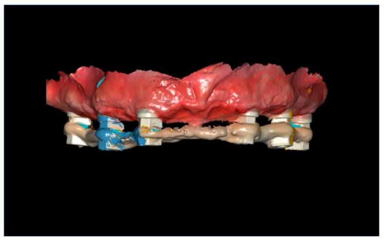
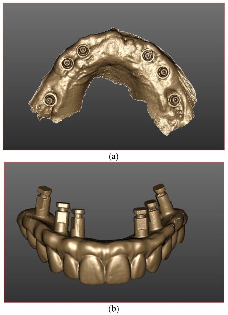
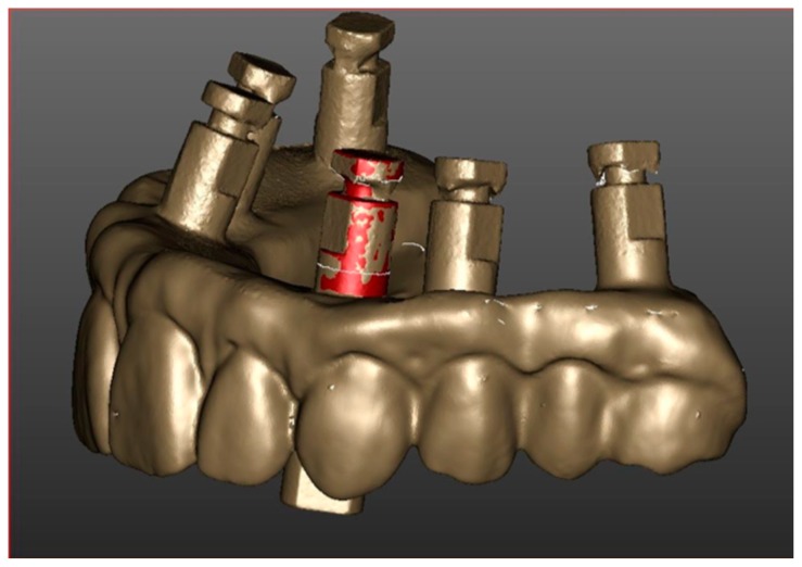
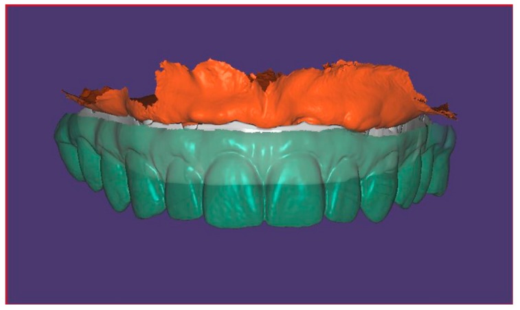
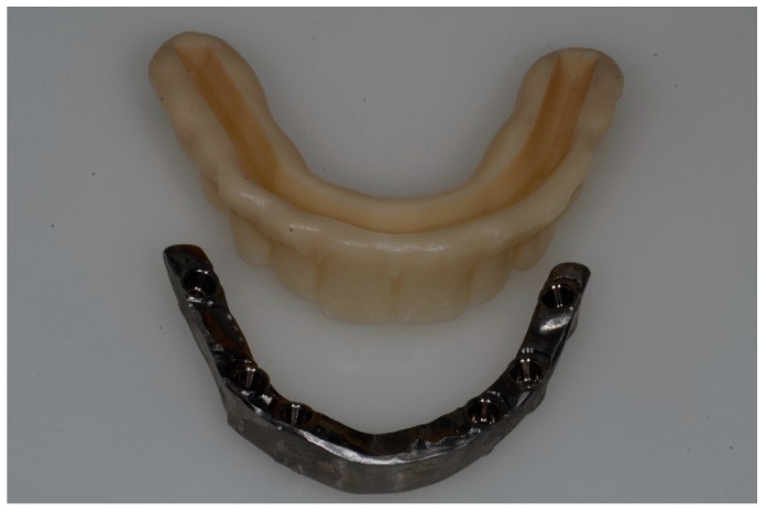
: The objective of this study was to compare conventional versus digital impressions for Full-Arch maxillary rehabilitations. : Patients selected for this study were treated with full-arch screw-retained rehabilitations supported by six immediately loaded dental implants. Patients have been scheduled randomly into control (conventional impression group, CIG) and test (digital impression group, DIG) groups respectively for a fully conventional workflow and a fully digital workflow. In both groups, within 24 h, temporary prostheses were delivered. Four months after the implant positioning, the two groups dealt with the fabrication of definitive restorations: conventional pick-up was performed in the control group, and definitive digital impressions were carried out in the test group. The time involved following these two procedures was recorded. Patients underwent intraoral digital radiographs to evaluate the accuracy of the framework-implant connection, check for the presence of voids at the bar-implant connection and measure bone level. Criteria used to assess success at the prosthetic level were the occurrence of prosthetic maintenance, the absence of fractures of the acrylic resin superstructure and voids. : A total of 50 patients received immediately loaded prostheses supported by six implants (total 300 implants). A fixture and prosthetic survival rate of 100% was observed. All digital X-ray examinations revealed a bar-implant connection accuracy and no voids. Differences that were not statistically significant ( > 0.05) in marginal bone loss were found between control and test groups. Significantly less time was spent to perform digital impression procedure ( < 0.05). : Clinical and radiological results of the test group advocate a satisfactory accuracy and predictability of the intraoral scanner (IOS) to be a reliable alternative in clinical practice for implant full-arch rehabilitations and suggest fabrication of definitive restorations with a successful marginal fit precision.
: 本研究的目的是比较全上颌修复的传统与数字化印模。 : 选择接受全上颌螺钉固位修复的患者,由六颗即刻负重种植体支持。患者随机分为对照组(传统印模组,CIG)和试验组(数字化印模组,DIG),分别采用全传统流程和全数字化流程。在两组中,均在 24 小时内交付临时修复体。种植体定位后 4 个月,两组分别处理最终修复体的制作:对照组进行常规取模,试验组进行最终数字化印模。记录这两个步骤所涉及的时间。患者接受口腔内数字化射线照相术,以评估框架-种植体连接的准确性,检查杆-种植体连接处是否有空隙,并测量骨水平。用于评估修复体水平成功的标准是出现修复体维护、丙烯酸树脂上层结构无断裂和无空隙。 : 共有 50 名患者接受了由六颗种植体(共 300 颗种植体)支持的即刻负重修复体。观察到固定器和修复体的存活率为 100%。所有数字射线照相检查均显示杆-种植体连接准确,无空隙。对照组和试验组之间的边缘骨丧失差异无统计学意义(> 0.05)。数字化印模程序的时间明显缩短(< 0.05)。 : 试验组的临床和放射学结果表明,口腔内扫描仪(IOS)具有令人满意的准确性和可预测性,可以作为临床实践中全颌种植体修复的可靠替代方法,并建议使用具有成功边缘拟合精度的最终修复体进行制作。