State Key Laboratory of Bioreactor Engineering, East China University of Science and Technology, 130 Meilong Rd, Shanghai, 200237, China.
Stem Cell Res Ther. 2019 Mar 13;10(1):91. doi: 10.1186/s13287-019-1181-5.
Adipose-derived mesenchymal stem cells (ADMSCs) are considered an efficient and important candidate for male infertility treatment because they contain pluripotent stem cells, which can differentiate into all cells from three germ layers. However, the efficient generation of male germ-like cell (MGLCs) is one of the key issues, and little is known about the mechanisms underlying generation of MGLCs. Herein, we attempt to improve the efficient generation of MGLCs derived during co-culturing of rat ADMSCs with SCs under retinoic acid (RA) and testosterone (T) treatment.
ADMSCs isolated from male SD rat were induced into generation of MGLCs by using respective methods in vitro. Transwell insert system was used for co-culturing. Busulfan-induced non-obstructive azoospermia rat mode was used to evaluate spermatogenic recovery ability of treated ADMSCs. Besides, the relative gene expression level was detected by reverse transcription PCR, quantitative RT-PCR. The relative protein expression level was detected by western blot (WB) and immunostaining analysis.
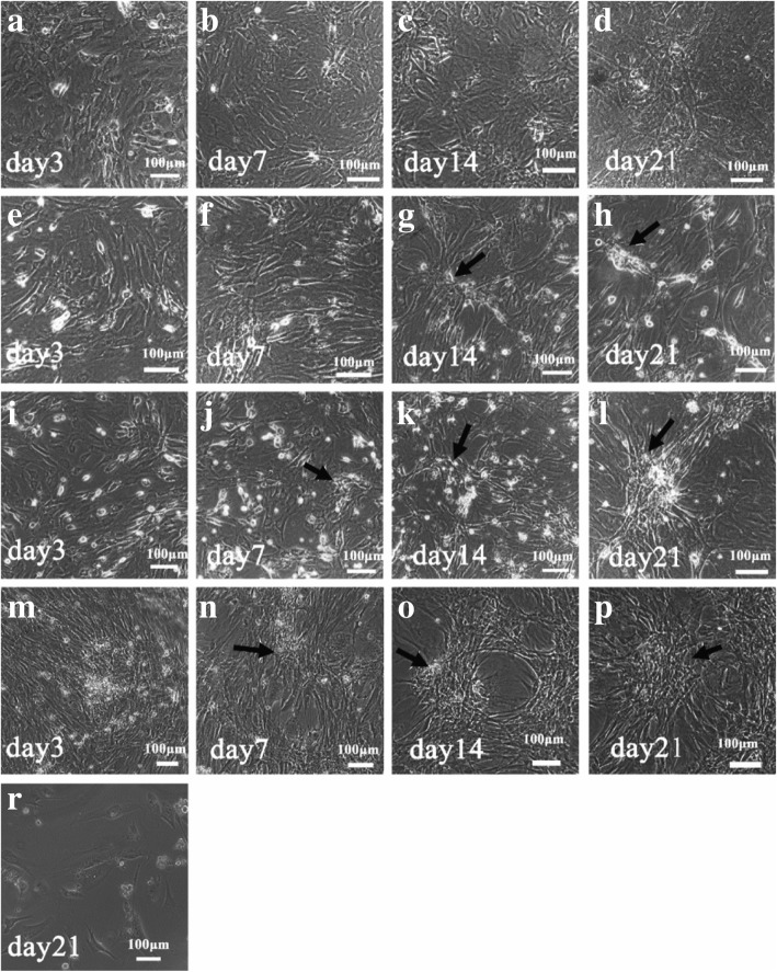
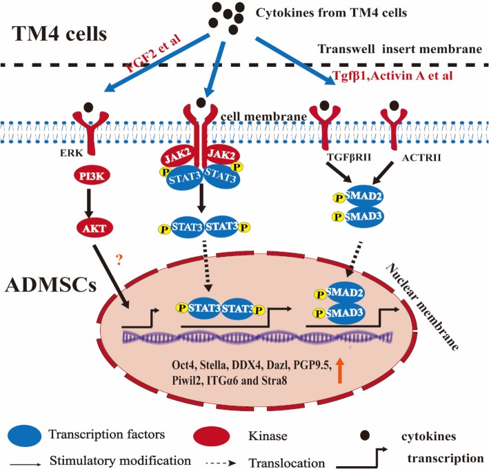
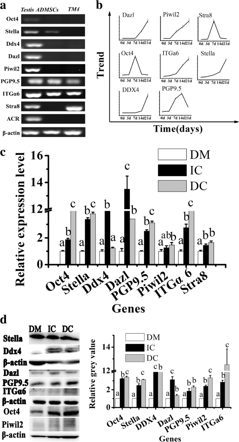
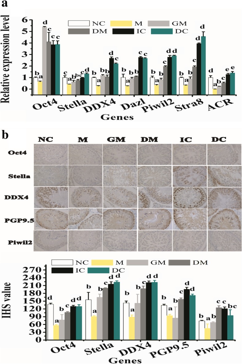
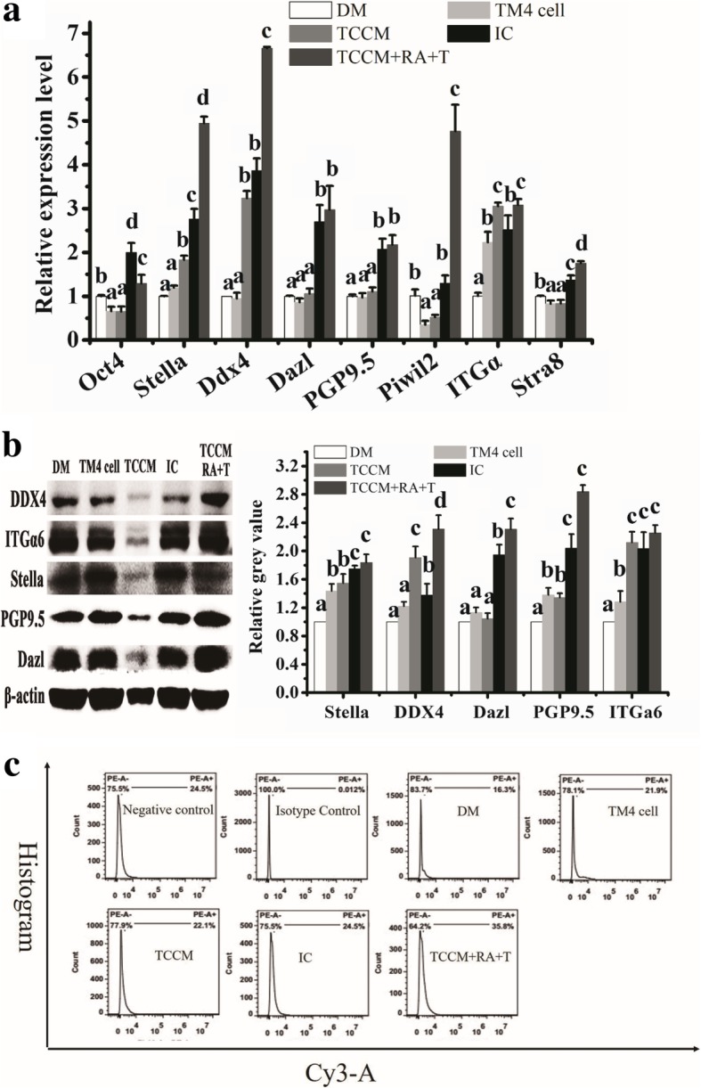
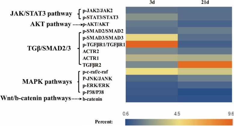
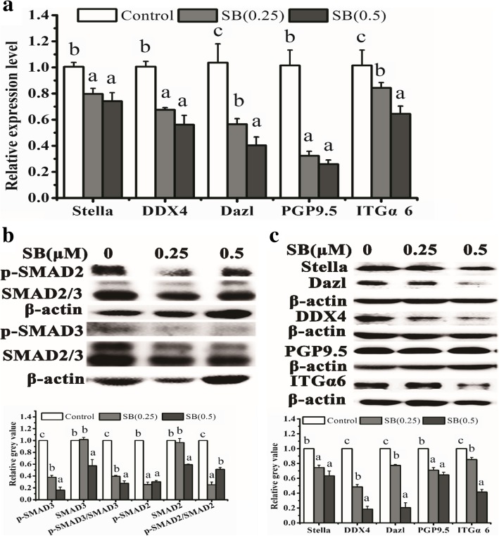
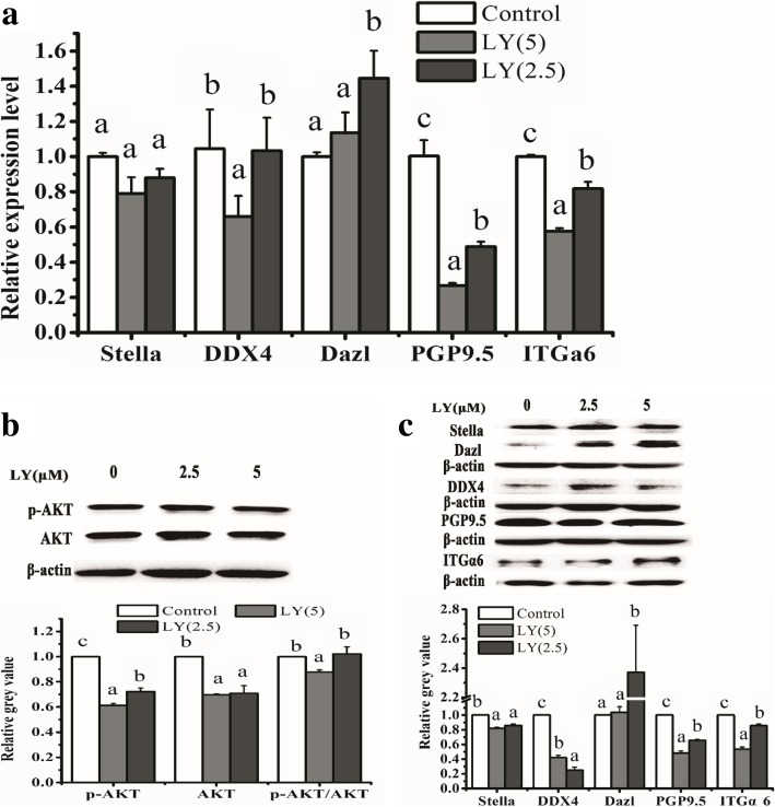
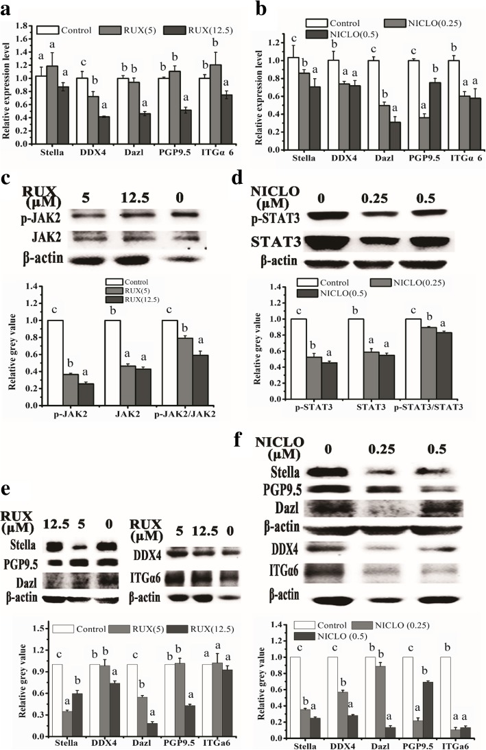
The results showed that ADMSCs co-cultured with TM4 cells under RA and T induction enhanced the formation of bigger and tightly packed MGLCs feature colonies in vitro. Moreover, the expression of male germ cell-related markers (Oct4, Stella, Ddx4, Dazl, PGP9.5, Stra8, and ITGα6) is significantly upregulated in TM4 cell-co-cultured ADMSCs in vitro and in busulfan-treated rat testis after injecting TM4 cell-treated ADMSCs for 2 months. Comparatively, the ADMSCs treated by TM4 cell with RA and T exhibited the highest expression of male germ cell-related markers. RA- and T-treated TM4 cell showed fewer dead cells and higher cytokine secretion than untreated groups. The protein expression level of TGFβ-SMAD2/3, JAK2-STAT3, and AKT pathways in ADMSCs co-cultured with TM4 cells under RA and T was higher than others. Whereas, downregulation of male germ cell-related marker expression subsequently inhibited the phosphorylation of SMAD2/3, JAK2, STAT3, and AKT.
These results suggested that TM4 cells could efficiently stimulate in vitro generation of MGLCs during co-culturing of ADMSCs under RA and T treatment. Conclusively, the ADMSCs co-cultured with TM4 cell under RA and T induction stimulate the efficient generation of MGLCs in vitro through activating TGFβ-SMAD2/3, JAK2-STAT3, and AKT pathways. Among them, JAK2-STAT3 and AKT pathways are being first reported to show involvement of in vitro generation of MGLCs during ADMSC co-culturing with SCs.
脂肪间充质干细胞(ADMSCs)被认为是男性不育治疗的有效且重要的候选物,因为它们含有多能干细胞,可分化为三个胚层的所有细胞。然而,高效地生成类精子细胞(MGLCs)是关键问题之一,目前对于生成 MGLCs 的机制知之甚少。在此,我们尝试通过在 RA 和 T 处理下将大鼠 ADMSCs 与 SC 共培养来提高 MGLCs 的有效生成。
从雄性 SD 大鼠中分离 ADMSCs,通过体外各自的方法诱导生成 MGLCs。使用 Transwell 插入系统进行共培养。采用布硫磷诱导的非梗阻性无精子症大鼠模型评估经处理的 ADMSCs 的生精恢复能力。此外,通过逆转录 PCR、定量 RT-PCR 检测相对基因表达水平,通过 Western blot(WB)和免疫染色分析检测相对蛋白表达水平。
结果表明,ADMSCs 在 RA 和 T 诱导下与 TM4 细胞共培养可增强体外更大且更紧密的 MGLC 特征集落的形成。此外,在体外 TM4 细胞共培养的 ADMSCs 中,以及在注射 TM4 细胞处理的 ADMSCs 2 个月后,在布硫磷处理的大鼠睾丸中,雄性生殖细胞相关标志物(Oct4、Stella、Ddx4、Dazl、PGP9.5、Stra8 和 ITGα6)的表达显著上调。相比之下,经 TM4 细胞处理的 ADMSCs 用 RA 和 T 处理后,雄性生殖细胞相关标志物的表达最高。RA 和 T 处理的 TM4 细胞表现出比未处理组更少的死亡细胞和更高的细胞因子分泌。在 RA 和 T 处理下与 TM4 细胞共培养的 ADMSCs 中,TGFβ-SMAD2/3、JAK2-STAT3 和 AKT 通路的蛋白表达水平高于其他通路。然而,下调雄性生殖细胞相关标志物的表达随后抑制了 SMAD2/3、JAK2、STAT3 和 AKT 的磷酸化。
这些结果表明,TM4 细胞可在 RA 和 T 处理下 ADMSCs 共培养过程中有效刺激体外 MGLC 的生成。总之,在 RA 和 T 诱导下与 TM4 细胞共培养的 ADMSCs 通过激活 TGFβ-SMAD2/3、JAK2-STAT3 和 AKT 通路来刺激体外 MGLC 的高效生成。其中,JAK2-STAT3 和 AKT 通路是首次报道在 ADMSC 与 SC 共培养过程中参与 MGLC 的体外生成。