University Clinical Center, University Hospital Medical University of Silesia, Katowice, Poland,
Department of Sexuology, Woman's Health Institute, School of Health Sciences in Katowice, Medical University of Silesia, Katowice, Poland.
Clin Interv Aging. 2019 Mar 8;14:505-514. doi: 10.2147/CIA.S189417. eCollection 2019.
To present optical coherence tomography (OCT) angiography features in patients with idiopathic full-thickness macular hole before and after vitrectomy.
Prospective case series study.
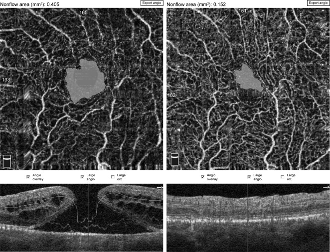
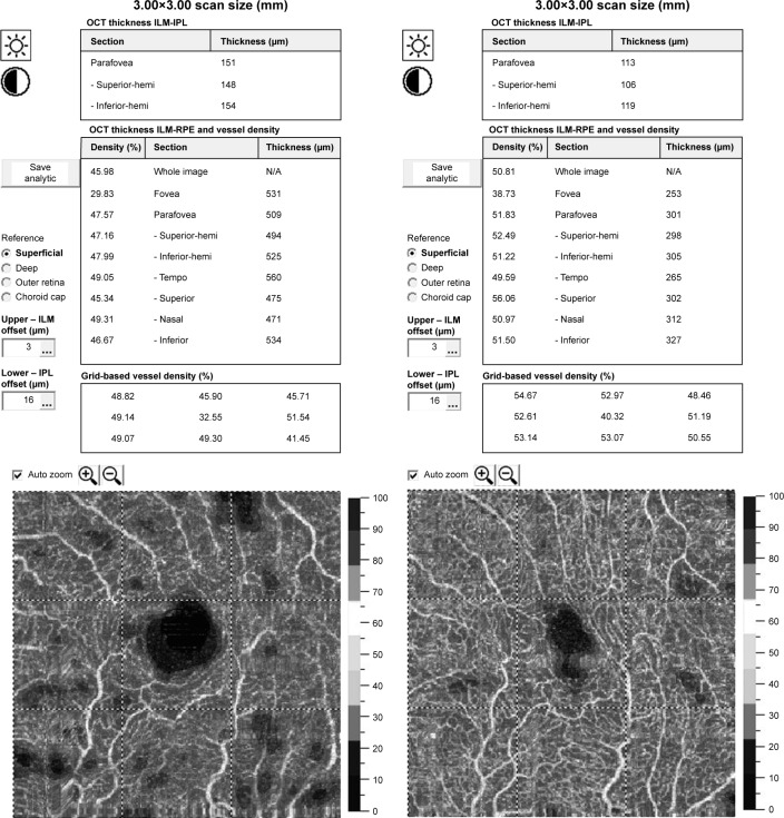
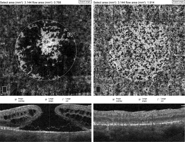
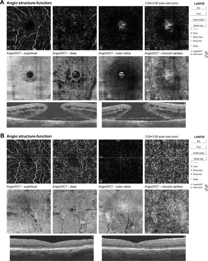
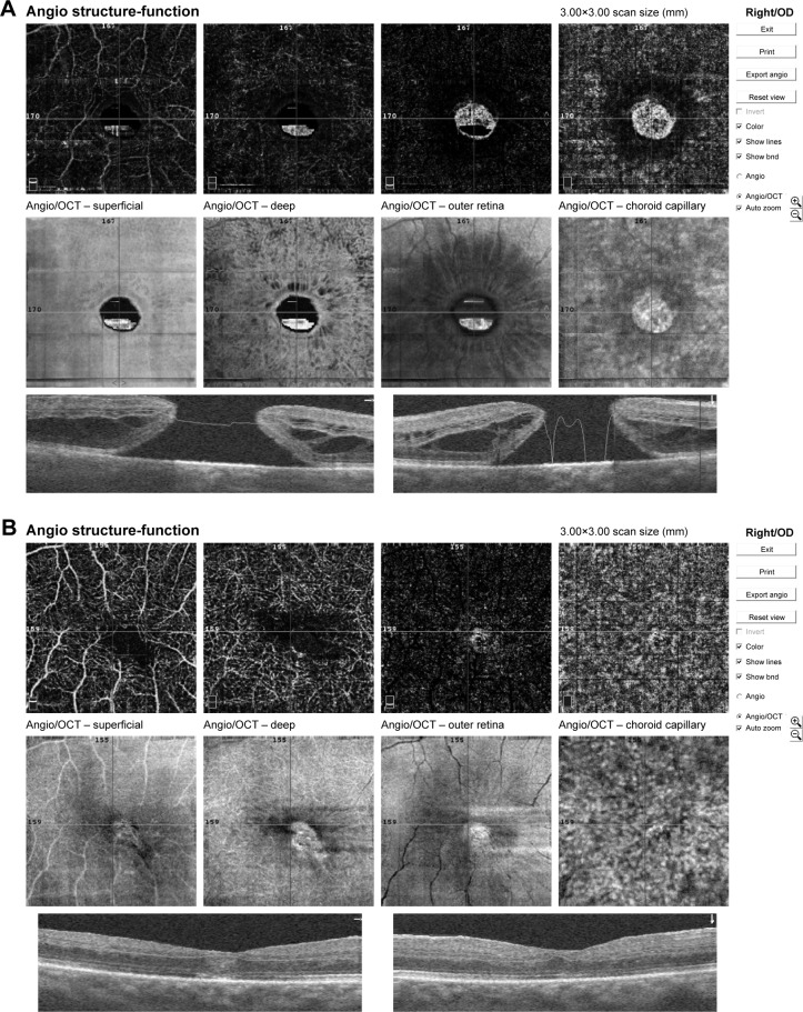
Patients presenting with an idiopathic full-thickness macular hole (IMH) who underwent posterior vitrectomy with internal limiting membrane peeling and gas tamponade were included in the study. En face OCT and OCT angiography (OCTA) was performed pre- and postoperatively using 3×3 mm scans (Optovue, XR Avanti). Foveal avascular zone (FAZ) area, macular hole size (MHS), central retinal thickness (CRT), macular parafoveal choriocapillary flow area (MCFA), and fovea vessel density (FVDS) were measured and assessed using OCTA. Best-corrected visual acuity (BCVA) was examined before and 3 months after surgery.
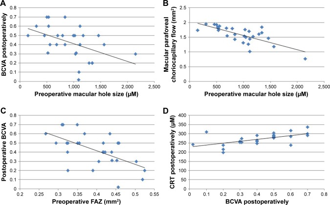
Twenty-eight eyes of 28 patients were included in the study. The mean age of patient group was 68.28 years. The hole was closed in all eyes after the initial surgery. OCTA showed enlargement of FAZ and increased CRT in foveal area. Mean preoperative FAZ area was 0.39±0.07 mm. En face images of the middle retina showed a range of preoperative cystic patterns surrounding the hole. BCVA was improved from 0.1±0.11 preoperatively to 0.42±0.17 postoperatively. Mean FAZ area was reduced to 0.24±0.07 mm postoperatively with resolution of macular hole and adjacent cystic areas. Mean CRT was reduced from 396±62.6 µm pre-operatively to 272±30.7 µm postoperatively. After vitrectomy, the parafoveal choriocapillary flow area and FVDS of IMH eyes increased compared with the preoperative measurements.
Quantitative evaluation of vascular and morphological changes following IMH surgery using OCTA shows the potential for recovery due to vascular and neuronal plasticity. OCTA showing vascular changes and their quantitative characteristics might be a useful tool for the assessment of macular holes before and after surgical treatment.
介绍特发性全层黄斑裂孔(IMH)患者玻璃体切除术前及术后的光学相干断层扫描血管造影(OCTA)特征。
前瞻性病例系列研究。
本研究纳入了接受玻璃体切除联合内界膜剥除和气体填充的特发性全层黄斑裂孔患者。使用 3×3mm 扫描(Optovue,XR Avanti)行术前和术后的黄斑区 OCT 和 OCTA。使用 OCTA 测量和评估黄斑中心凹无血管区(FAZ)面积、黄斑裂孔大小(MHS)、中心视网膜厚度(CRT)、黄斑旁中心凹脉络膜血流面积(MCFA)和黄斑中心凹血管密度(FVDS)。手术前和手术后 3 个月检查最佳矫正视力(BCVA)。
本研究纳入了 28 例 28 只眼。患者组的平均年龄为 68.28 岁。所有患者的裂孔在初次手术后均闭合。OCTA 显示 FAZ 扩大,黄斑中心凹 CRT 增加。术前 FAZ 平均面积为 0.39±0.07mm。中视网膜的 OCT 图像显示术前存在各种围绕裂孔的囊样形态。术前 BCVA 为 0.1±0.11,术后提高至 0.42±0.17。术后黄斑裂孔及相邻囊腔区消失,平均 FAZ 面积缩小至 0.24±0.07mm。术前 CRT 为 396±62.6μm,术后降至 272±30.7μm。玻璃体切除术后,IMH 眼的旁中心凹脉络膜血流面积和 FVDS 较术前增加。
使用 OCTA 对 IMH 手术后的血管和形态变化进行定量评估,显示了血管和神经元可塑性恢复的潜力。OCTA 显示血管变化及其定量特征可能成为黄斑裂孔手术前后评估的有用工具。