Laboratory of Biophysics of Excitable Systems, Moscow Institute of Physics and Technology, Dolgoprudny, Russia.
Department of Physics and Astronomy, Ghent University, Ghent, Belgium.
PLoS Comput Biol. 2019 Mar 18;15(3):e1006597. doi: 10.1371/journal.pcbi.1006597. eCollection 2019 Mar.
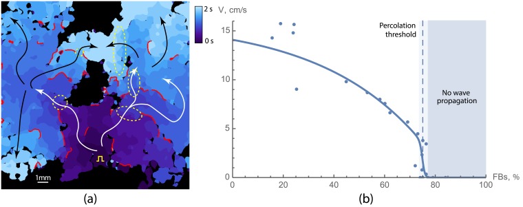
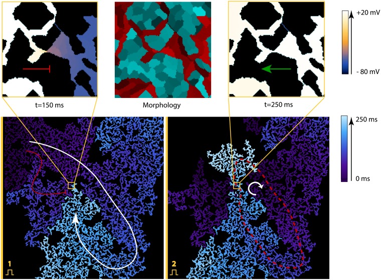
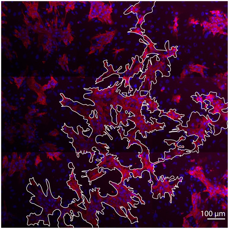
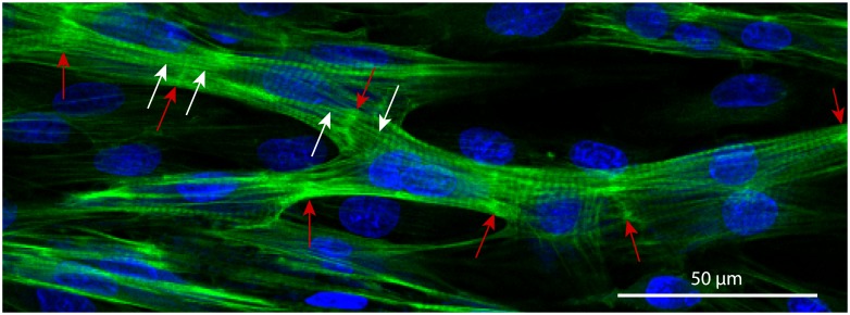
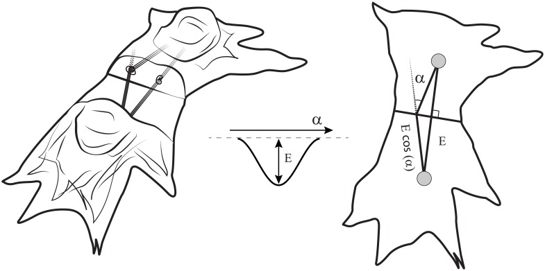
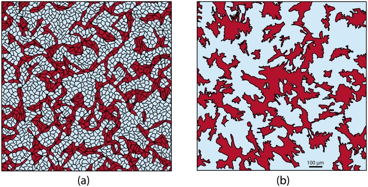
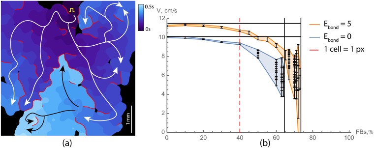
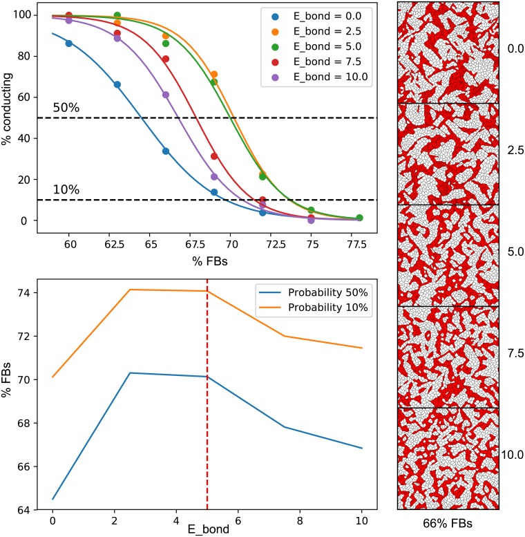
Cardiac fibrosis occurs in many forms of heart disease and is considered to be one of the main arrhythmogenic factors. Regions with a high density of fibroblasts are likely to cause blocks of wave propagation that give rise to dangerous cardiac arrhythmias. Therefore, studies of the wave propagation through these regions are very important, yet the precise mechanisms leading to arrhythmia formation in fibrotic cardiac tissue remain poorly understood. Particularly, it is not clear how wave propagation is organized at the cellular level, as experiments show that the regions with a high percentage of fibroblasts (65-75%) are still conducting electrical signals, whereas geometric analysis of randomly distributed conducting and non-conducting cells predicts connectivity loss at 40% at the most (percolation threshold). To address this question, we used a joint in vitro-in silico approach, which combined experiments in neonatal rat cardiac monolayers with morphological and electrophysiological computer simulations. We have shown that the main reason for sustainable wave propagation in highly fibrotic samples is the formation of a branching network of cardiomyocytes. We have successfully reproduced the morphology of conductive pathways in computer modelling, assuming that cardiomyocytes align their cytoskeletons to fuse into cardiac syncytium. The electrophysiological properties of the monolayers, such as conduction velocity, conduction blocks and wave fractionation, were reproduced as well. In a virtual cardiac tissue, we have also examined the wave propagation at the subcellular level, detected wavebreaks formation and its relation to the structure of fibrosis and, thus, analysed the processes leading to the onset of arrhythmias.
心肌纤维化存在于多种形式的心脏病中,被认为是主要的心律失常因素之一。成纤维细胞密度较高的区域可能导致波传播受阻,从而引发危险的心律失常。因此,研究这些区域的波传播非常重要,但纤维化心肌组织中导致心律失常形成的确切机制仍知之甚少。特别是,目前还不清楚波传播在细胞水平上是如何组织的,因为实验表明,纤维化区域(65-75%)仍在传导电信号,而随机分布的传导和非传导细胞的几何分析预测,在最多 40%(渗流阈值)的情况下,连接就会丢失。为了解决这个问题,我们采用了一种体外与计算机模拟相结合的联合方法,结合了新生大鼠心脏单层的实验和形态学及电生理学计算机模拟。我们表明,在高度纤维化的样本中可持续传播波的主要原因是形成了分支状的心肌细胞网络。我们成功地在计算机建模中再现了传导通路的形态,假设心肌细胞调整其细胞骨架以融合成心脏合胞体。单层的电生理特性,如传导速度、传导阻滞和波分裂,也得到了再现。在虚拟心脏组织中,我们还在亚细胞水平上检查了波传播,检测到波破裂的形成及其与纤维化结构的关系,从而分析了导致心律失常发生的过程。