• 文献检索
  • 文档翻译
  • 深度研究
  • 学术资讯
  • Suppr Zotero 插件Zotero 插件
  • 邀请有礼
  • 套餐&价格
  • 历史记录
应用&插件
Suppr Zotero 插件Zotero 插件浏览器插件Mac 客户端Windows 客户端微信小程序
定价
高级版会员购买积分包购买API积分包
服务
文献检索文档翻译深度研究API 文档MCP 服务
关于我们
关于 Suppr公司介绍联系我们用户协议隐私条款
关注我们

Suppr 超能文献

核心技术专利:CN118964589B侵权必究
粤ICP备2023148730 号-1Suppr @ 2026

文献检索

告别复杂PubMed语法,用中文像聊天一样搜索,搜遍4000万医学文献。AI智能推荐,让科研检索更轻松。

立即免费搜索

文件翻译

保留排版,准确专业,支持PDF/Word/PPT等文件格式,支持 12+语言互译。

免费翻译文档

深度研究

AI帮你快速写综述,25分钟生成高质量综述,智能提取关键信息,辅助科研写作。

立即免费体验

在肥厚型心肌病患者中,正电子发射断层扫描(PET)测量的心肌血流储备与超声心动图估测的肺动脉收缩压的相关性。

Association of PET-measured myocardial flow reserve with echocardiography-estimated pulmonary artery systolic pressure in patients with hypertrophic cardiomyopathy.

机构信息

Department of Nuclear Medicine, Xiangya Hospital, Central South University, Changsha, China.

The Russell H. Morgan Department of Radiology and Radiological Science, Johns Hopkins School of Medicine, Baltimore, Maryland, United States of America.

出版信息

PLoS One. 2019 Mar 20;14(3):e0212573. doi: 10.1371/journal.pone.0212573. eCollection 2019.

DOI:10.1371/journal.pone.0212573
PMID:30893304
原文链接:https://pmc.ncbi.nlm.nih.gov/articles/PMC6426216/
Abstract

BACKGROUND

Pulmonary hypertension (PH) is a known complication of HCM and is a strong predictor of mortality. We aim to investigate the relationship between microvascular dysfunction measured by quantitative PET and PH in HCM patients.

METHODS

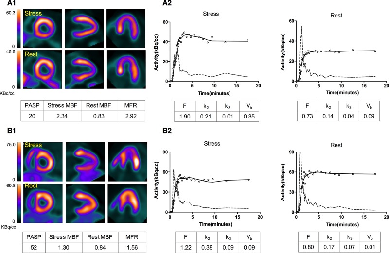
Eighty-nine symptomatic HCM patients were included in the study. Each patient underwent two 20-min 13N-NH3 dynamic PET scans for rest and stress conditions, respectively. A 2-tissue irreversible compartmental model was used to fit the segments time activity curves for estimating segmental and global myocardial blood flow (MBF) and myocardial flow reserve (MFR). Echocardiographic derived PASP was utilized to estimate PH.

RESULTS

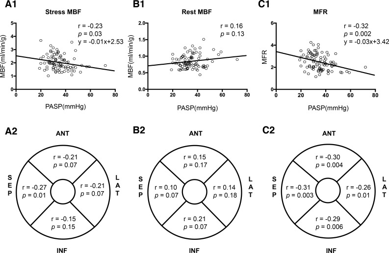
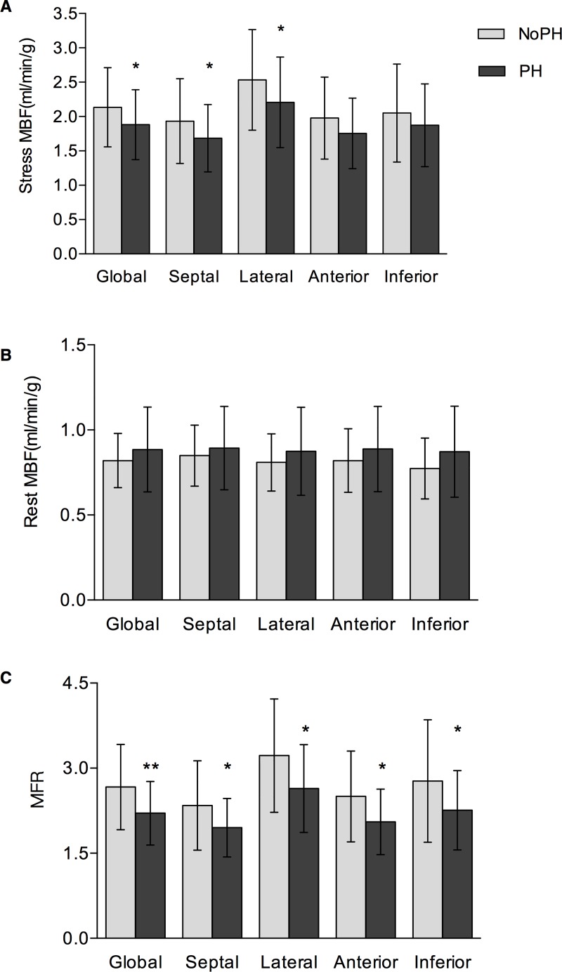
Patients were categorized into two groups across PASP: PH (PASP > 36 mmHg) and no-PH (PASP ≤ 36 mmHg). patients with PH had larger left atrium, ratio of higher inflow early diastole (E) and atrial contraction (A) waves, E/A, and ratio of inflow and peak early diastolic waves, E/e', significantly reduced global stress MBF (1.85 ± 0.52 vs. 2.13 ± 0.56 ml/min/g; p = 0.024) and MFR (2.21 ± 0.57 vs. 2.62 ± 0.75; p = 0.005), while the MBFs at rest between the two groups were similar. There were significant negative correlations between global stress MBF/MFR and PASP (stress MBF: r = -0.23, p = 0.03; MFR: r = -0.32, p = 0.002); for regional MBF and MFR measurements, the highest linear correlation coefficients were observed in the septal wall (stress MBF: r = -0.27, p = 0.01; MFR: r = -0.31, p = 0.003). Global MFR was identified to be independent predictor for PH in multivariate regression analysis.

CONCLUSION

Echocardiography-derived PASP is negatively correlated with global MFR measured by 13N-NH3 dynamic PET. Global MFR is suggested to be an index of PH in HCM patients.

摘要

背景

肺动脉高压(PH)是肥厚型心肌病(HCM)的已知并发症,也是死亡率的强有力预测指标。我们旨在研究通过定量 PET 测量的微血管功能障碍与 HCM 患者 PH 之间的关系。

方法

研究纳入 89 例有症状的 HCM 患者。每位患者分别进行两次 20 分钟 13N-NH3 动态 PET 扫描,分别用于静息和应激条件。使用 2 组织不可逆房室模型拟合节段时间活动曲线,以估计节段和整体心肌血流(MBF)和心肌血流储备(MFR)。超声心动图衍生的 PASP 用于估计 PH。

结果

患者根据 PASP 分为两组:PH(PASP > 36mmHg)和非 PH(PASP ≤ 36mmHg)。PH 患者的左心房较大,心房收缩(A)波与早期舒张(E)波的比值较高,E/A 和流入和峰值早期舒张波的比值,E/e',静息时两组之间的整体应激 MBF(1.85 ± 0.52 vs. 2.13 ± 0.56ml/min/g;p = 0.024)和 MFR(2.21 ± 0.57 vs. 2.62 ± 0.75;p = 0.005)明显降低,而两组之间的静息 MBF 相似。全球应激 MBF/MFR 与 PASP 呈显著负相关(应激 MBF:r = -0.23,p = 0.03;MFR:r = -0.32,p = 0.002);对于区域性 MBF 和 MFR 测量,在间隔壁上观察到最高的线性相关系数(应激 MBF:r = -0.27,p = 0.01;MFR:r = -0.31,p = 0.003)。多元回归分析显示,全球 MFR 是 PH 的独立预测因子。

结论

超声心动图衍生的 PASP 与 13N-NH3 动态 PET 测量的整体 MFR 呈负相关。整体 MFR 被认为是 HCM 患者 PH 的一个指标。

https://cdn.ncbi.nlm.nih.gov/pmc/blobs/0c7d/6426216/52a4d1ade451/pone.0212573.g003.jpg
https://cdn.ncbi.nlm.nih.gov/pmc/blobs/0c7d/6426216/11c75c2d6e9a/pone.0212573.g001.jpg
https://cdn.ncbi.nlm.nih.gov/pmc/blobs/0c7d/6426216/ca670f766550/pone.0212573.g002.jpg
https://cdn.ncbi.nlm.nih.gov/pmc/blobs/0c7d/6426216/52a4d1ade451/pone.0212573.g003.jpg
https://cdn.ncbi.nlm.nih.gov/pmc/blobs/0c7d/6426216/11c75c2d6e9a/pone.0212573.g001.jpg
https://cdn.ncbi.nlm.nih.gov/pmc/blobs/0c7d/6426216/ca670f766550/pone.0212573.g002.jpg
https://cdn.ncbi.nlm.nih.gov/pmc/blobs/0c7d/6426216/52a4d1ade451/pone.0212573.g003.jpg

相似文献

1
Association of PET-measured myocardial flow reserve with echocardiography-estimated pulmonary artery systolic pressure in patients with hypertrophic cardiomyopathy.在肥厚型心肌病患者中,正电子发射断层扫描(PET)测量的心肌血流储备与超声心动图估测的肺动脉收缩压的相关性。
PLoS One. 2019 Mar 20;14(3):e0212573. doi: 10.1371/journal.pone.0212573. eCollection 2019.
2
Comparison of two software systems for quantification of myocardial blood flow in patients with hypertrophic cardiomyopathy.肥厚型心肌病患者心肌血流定量的两种软件系统比较
J Nucl Cardiol. 2019 Aug;26(4):1243-1253. doi: 10.1007/s12350-017-1155-x. Epub 2018 Jan 22.
3
Assessment of intravascular and extravascular mechanisms of myocardial perfusion abnormalities in obstructive hypertrophic cardiomyopathy by myocardial contrast echocardiography.应用心肌对比超声心动图评估梗阻性肥厚型心肌病中心肌灌注异常的血管内和血管外机制。
Heart. 2007 Oct;93(10):1204-12. doi: 10.1136/hrt.2006.110460. Epub 2007 May 8.
4
Comparison between quantitative cardiac magnetic resonance perfusion imaging and [O]HO positron emission tomography.定量心脏磁共振灌注成像与 [O]HO 正电子发射断层扫描比较。
Eur J Nucl Med Mol Imaging. 2020 Jul;47(7):1688-1697. doi: 10.1007/s00259-019-04641-9. Epub 2019 Dec 10.
5
Diagnostic value of PET-measured longitudinal flow gradient for the identification of coronary artery disease.PET 测量的纵向血流梯度对冠状动脉疾病的诊断价值。
JACC Cardiovasc Imaging. 2014 Apr;7(4):387-96. doi: 10.1016/j.jcmg.2014.01.001. Epub 2014 Mar 13.
6
Association of Myocardial Blood Flow Reserve With Adverse Left Ventricular Remodeling in Patients With Aortic Stenosis: The Microvascular Disease in Aortic Stenosis (MIDAS) Study.主动脉瓣狭窄患者心肌血流储备与不良左心室重构的相关性:主动脉瓣狭窄中的微血管疾病(MIDAS)研究。
JAMA Cardiol. 2022 Jan 1;7(1):93-99. doi: 10.1001/jamacardio.2021.3396.
7
Comparison of clinical tools for measurements of regional stress and rest myocardial blood flow assessed with 13N-ammonia PET/CT.比较采用 13N-氨 PET/CT 评估的局部应力和静息心肌血流的临床工具。
J Nucl Med. 2012 Feb;53(2):171-81. doi: 10.2967/jnumed.111.095398. Epub 2012 Jan 6.
8
Quantification of myocardial blood flow (MBF) and reserve (MFR) incorporated with a novel segmentation approach: Assessments of quantitative precision and the lower limit of normal MBF and MFR in patients.采用新型分段方法对心肌血流(MBF)和储备(MFR)进行定量评估:在患者中评估定量精度和正常 MBF 和 MFR 的下限。
J Nucl Cardiol. 2021 Aug;28(4):1236-1248. doi: 10.1007/s12350-020-02278-y. Epub 2020 Jul 26.
9
PET/CT assessment of symptomatic individuals with obstructive and nonobstructive hypertrophic cardiomyopathy.PET/CT 评估梗阻性和非梗阻性肥厚型心肌病有症状患者。
J Nucl Med. 2012 Mar;53(3):407-14. doi: 10.2967/jnumed.111.096156. Epub 2012 Feb 7.
10
Optimization of SPECT Measurement of Myocardial Blood Flow with Corrections for Attenuation, Motion, and Blood Binding Compared with PET.与PET相比,通过衰减、运动和血液结合校正优化心肌血流的SPECT测量
J Nucl Med. 2017 Dec;58(12):2013-2019. doi: 10.2967/jnumed.117.191049. Epub 2017 Jun 13.

引用本文的文献

1
The Utility of Nuclear Imaging in Hypertrophic Cardiomyopathy: A Narrative Review.核成像在肥厚型心肌病中的应用:一篇叙述性综述
J Clin Med. 2025 Mar 22;14(7):2183. doi: 10.3390/jcm14072183.
2
Microvascular Dysfunction as a Systemic Disease: A Review of the Evidence.微血管功能障碍作为一种全身性疾病:证据综述。
Am J Med. 2022 Sep;135(9):1059-1068. doi: 10.1016/j.amjmed.2022.04.006. Epub 2022 Apr 23.

本文引用的文献

1
Higher incidence of vasodilator-induced left ventricular cavity dilation by PET when compared to treadmill exercise-ECHO in hypertrophic cardiomyopathy.与平板运动超声心动图相比,正电子发射断层扫描(PET)在肥厚型心肌病中显示出更高的血管扩张剂诱导的左心室腔扩张发生率。
J Nucl Cardiol. 2020 Dec;27(6):2031-2043. doi: 10.1007/s12350-018-01521-x. Epub 2018 Nov 19.
2
Pulmonary hypertension and clinical correlates in hypertrophic cardiomyopathy.肥厚型心肌病中的肺动脉高压与临床相关性。
Int J Cardiol. 2017 Dec 1;248:326-332. doi: 10.1016/j.ijcard.2017.07.010. Epub 2017 Jul 14.
3
Role of PET to evaluate coronary microvascular dysfunction in non-ischemic cardiomyopathies.
正电子发射断层扫描(PET)在评估非缺血性心肌病中冠状动脉微血管功能障碍的作用。
Heart Fail Rev. 2017 Jul;22(4):455-464. doi: 10.1007/s10741-017-9628-1.
4
Mechanism of Progressive Heart Failure and Significance of Pulmonary Hypertension in Obstructive Hypertrophic Cardiomyopathy.梗阻性肥厚型心肌病中进行性心力衰竭的机制及肺动脉高压的意义
Circ Heart Fail. 2017 Apr;10(4):e003689. doi: 10.1161/CIRCHEARTFAILURE.116.003689.
5
Myocardial blood flow and left ventricular functional reserve in hypertrophic cardiomyopathy: a NH gated PET study.肥厚型心肌病中心肌血流与左心室功能储备:一项氮-13 氨门控正电子发射断层扫描研究
Eur J Nucl Med Mol Imaging. 2017 May;44(5):866-875. doi: 10.1007/s00259-016-3603-2. Epub 2017 Jan 3.
6
Positron-emission tomography myocardial blood flow quantification in hypertrophic cardiomyopathy.肥厚型心肌病的正电子发射断层扫描心肌血流定量分析
Q J Nucl Med Mol Imaging. 2016 Dec;60(4):354-61. Epub 2016 Sep 9.
7
Pulmonary hypertension is associated with worse survival in hypertrophic cardiomyopathy.肺动脉高压与肥厚型心肌病患者较差的生存率相关。
Eur Heart J Cardiovasc Imaging. 2016 Jun;17(6):604-10. doi: 10.1093/ehjci/jew024. Epub 2016 Feb 27.
8
The pathophysiology of pulmonary hypertension in left heart disease.左心疾病中肺动脉高压的病理生理学
Am J Physiol Lung Cell Mol Physiol. 2015 Nov 1;309(9):L924-41. doi: 10.1152/ajplung.00146.2015. Epub 2015 Aug 21.
9
2015 ESC/ERS Guidelines for the diagnosis and treatment of pulmonary hypertension: The Joint Task Force for the Diagnosis and Treatment of Pulmonary Hypertension of the European Society of Cardiology (ESC) and the European Respiratory Society (ERS): Endorsed by: Association for European Paediatric and Congenital Cardiology (AEPC), International Society for Heart and Lung Transplantation (ISHLT).2015年欧洲心脏病学会(ESC)/欧洲呼吸学会(ERS)肺动脉高压诊断和治疗指南:欧洲心脏病学会(ESC)和欧洲呼吸学会(ERS)肺动脉高压诊断和治疗联合工作组:得到以下组织认可:欧洲儿科和先天性心脏病协会(AEPC)、国际心肺移植学会(ISHLT)。
Eur Heart J. 2016 Jan 1;37(1):67-119. doi: 10.1093/eurheartj/ehv317. Epub 2015 Aug 29.
10
Apparent left ventricular cavity dilatation during PET/CT in hypertrophic cardiomyopathy: Clinical predictors and potential mechanisms.肥厚型心肌病患者在PET/CT检查时出现的左心室腔明显扩张:临床预测因素及潜在机制
J Nucl Cardiol. 2016 Dec;23(6):1304-1314. doi: 10.1007/s12350-015-0158-8. Epub 2015 May 20.