Department of Radiology, Netherlands Cancer Institute, Amsterdam; GROW School of Oncology and Developmental Biology, Maastricht, The Netherlands; Departments of Radiation Oncology; Radiology, Dana Farber Cancer Institute, Brigham and Women's Hospital, Harvard Medical School, Boston, USA.
Department of Radiology, Netherlands Cancer Institute, Amsterdam; Department of Radiology, Milano-Bicocca University, San Gerardo Hospital, Monza, Italy.
Ann Oncol. 2019 Jun 1;30(6):998-1004. doi: 10.1093/annonc/mdz108.
Immunotherapy is regarded as one of the major breakthroughs in cancer treatment. Despite its success, only a subset of patients responds-urging the quest for predictive biomarkers. We hypothesize that artificial intelligence (AI) algorithms can automatically quantify radiographic characteristics that are related to and may therefore act as noninvasive radiomic biomarkers for immunotherapy response.
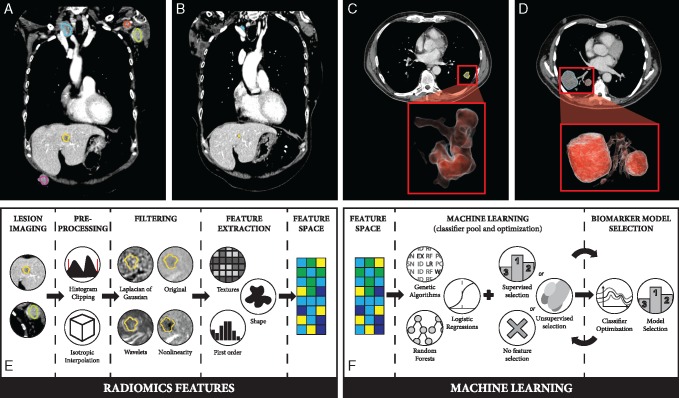
In this study, we analyzed 1055 primary and metastatic lesions from 203 patients with advanced melanoma and non-small-cell lung cancer (NSCLC) undergoing anti-PD1 therapy. We carried out an AI-based characterization of each lesion on the pretreatment contrast-enhanced CT imaging data to develop and validate a noninvasive machine learning biomarker capable of distinguishing between immunotherapy responding and nonresponding. To define the biological basis of the radiographic biomarker, we carried out gene set enrichment analysis in an independent dataset of 262 NSCLC patients.
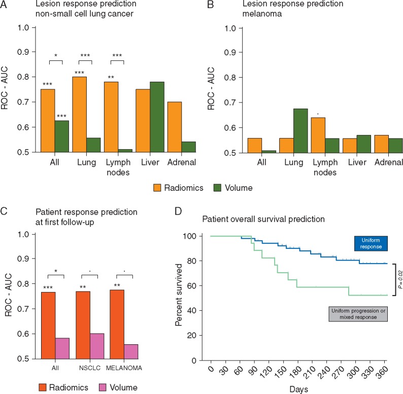
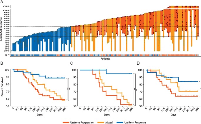
The biomarker reached significant performance on NSCLC lesions (up to 0.83 AUC, P < 0.001) and borderline significant for melanoma lymph nodes (0.64 AUC, P = 0.05). Combining these lesion-wide predictions on a patient level, immunotherapy response could be predicted with an AUC of up to 0.76 for both cancer types (P < 0.001), resulting in a 1-year survival difference of 24% (P = 0.02). We found highly significant associations with pathways involved in mitosis, indicating a relationship between increased proliferative potential and preferential response to immunotherapy.
These results indicate that radiographic characteristics of lesions on standard-of-care imaging may function as noninvasive biomarkers for response to immunotherapy, and may show utility for improved patient stratification in both neoadjuvant and palliative settings.
免疫疗法被认为是癌症治疗的重大突破之一。尽管取得了成功,但只有一部分患者有反应,这促使人们寻求预测生物标志物。我们假设人工智能 (AI) 算法可以自动量化与免疫治疗反应相关的影像学特征,这些特征可能成为非侵入性的放射组学生物标志物。
在这项研究中,我们分析了 203 名接受抗 PD-1 治疗的晚期黑色素瘤和非小细胞肺癌 (NSCLC) 患者的 1055 个原发和转移病灶。我们对每个病灶的预处理对比增强 CT 成像数据进行基于 AI 的特征描述,以开发和验证一种能够区分免疫治疗反应和非反应的非侵入性机器学习生物标志物。为了定义放射学生物标志物的生物学基础,我们在 262 名 NSCLC 患者的独立数据集上进行了基因集富集分析。
该生物标志物在 NSCLC 病灶上达到了显著的性能(最高 AUC 为 0.83,P<0.001),在黑色素瘤淋巴结上达到了边缘显著水平(0.64 AUC,P=0.05)。在患者层面上结合这些病灶级别的预测,免疫治疗反应可以通过两种癌症类型的 AUC 达到 0.76(P<0.001)进行预测,导致 1 年生存率差异为 24%(P=0.02)。我们发现与涉及有丝分裂的途径高度相关,表明增殖潜力增加与对免疫治疗的优先反应之间存在关系。
这些结果表明,标准治疗成像上的病灶影像学特征可能作为免疫治疗反应的非侵入性生物标志物,并可能在新辅助和姑息治疗中改善患者分层方面显示出实用性。