Neuroimaging Center, University Medical Center Groningen, University of Groningen, Neuroimaging Center 3111, Antonius Deusinglaan 2, 9713 AW Groningen, the Netherlands; Department of Clinical Neurophysiology and MEG Center, Amsterdam UMC, Vrije Universiteit Amsterdam, Neuroscience Campus Amsterdam, Postbus 7057, 1007 MB Amsterdam, the Netherlands; Department of Psychiatry, Brain Center Rudolf Magnus, University Medical Center Utrecht, Postbus 85500, 3508 GA Utrecht, the Netherlands.
Department of Neurology, St. Antonius Ziekenhuis, Nieuwegein, Utrecht, the Netherlands.
Neuroimage Clin. 2019;22:101752. doi: 10.1016/j.nicl.2019.101752. Epub 2019 Mar 13.
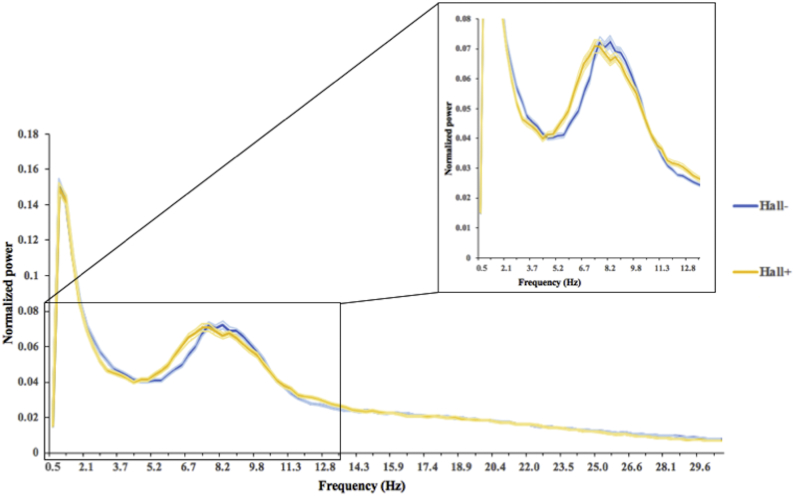
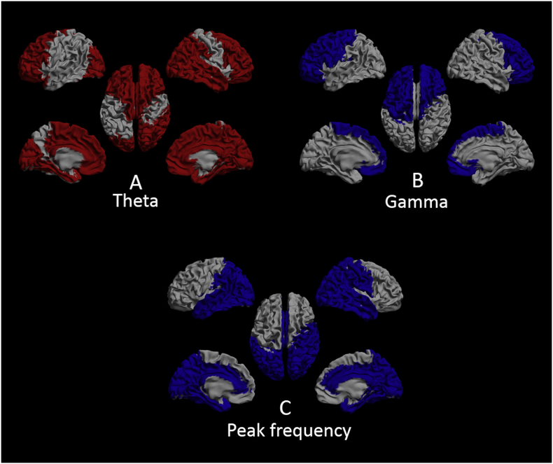
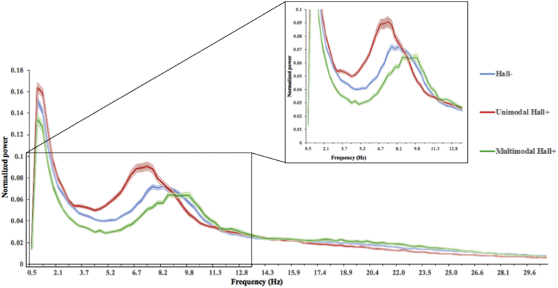
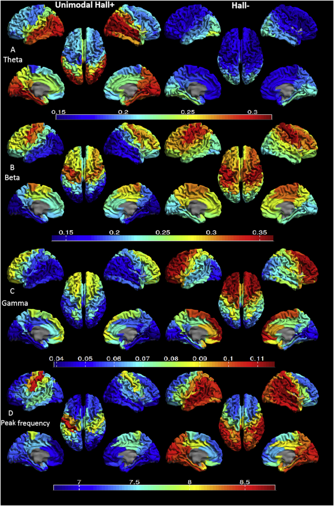
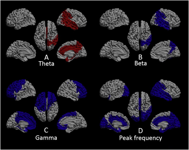
To gain insight into possible underlying mechanism(s) of visual hallucinations (VH) in Parkinson's disease (PD), we explored changes in local oscillatory activity in different frequency bands with source-space magnetoencephalography (MEG). Eyes-closed resting-state MEG recordings were obtained from 20 PD patients with hallucinations (Hall+) and 20 PD patients without hallucinations (Hall-), matched for age, gender and disease severity. The Hall+ group was subdivided into 10 patients with VH only (unimodal Hall+) and 10 patients with multimodal hallucinations (multimodal Hall+). Subsequently, neuronal activity at source-level was reconstructed using an atlas-based beamforming approach resulting in source-space time series for 78 cortical and 12 subcortical regions of interest in the automated anatomical labeling (AAL) atlas. Peak frequency (PF) and relative power in six frequency bands (delta, theta, alpha1, alpha2, beta and gamma) were compared between Hall+ and Hall-, unimodal Hall+ and Hall-, multimodal Hall+ and Hall-, and unimodal Hall+ and multimodal Hall+ patients. PF and relative power per frequency band did not differ between Hall+ and Hall-, and multimodal Hall+ and Hall- patients. Compared to the Hall- group, unimodal Hall+ patients showed significantly higher relative power in the theta band (p = 0.005), and significantly lower relative power in the beta (p = 0.029) and gamma (p = 0.007) band, and lower PF (p = 0.011). Compared to the unimodal Hall+, multimodal Hall+ showed significantly higher PF (p = 0.007). In conclusion, a subset of PD patients with only VH showed slowing of MEG-based resting-state brain activity with an increase in theta activity, and a concomitant decrease in beta and gamma activity, which could indicate central cholinergic dysfunction as underlying mechanism of VH in PD. This signature was absent in PD patients with multimodal hallucinations.
为了深入了解帕金森病(PD)中视觉幻觉(VH)的潜在机制,我们使用源空间脑磁图(MEG)探索了不同频带局部振荡活动的变化。从 20 名有幻觉(Hall+)的 PD 患者和 20 名无幻觉(Hall-)的 PD 患者中获得闭眼静息状态 MEG 记录,这些患者在年龄、性别和疾病严重程度上相匹配。Hall+组进一步分为 10 名仅有单模态幻觉(单模态 Hall+)的患者和 10 名有多模态幻觉(多模态 Hall+)的患者。随后,使用基于图谱的波束形成方法重建源水平的神经元活动,从而为自动解剖标记(AAL)图谱中的 78 个皮质和 12 个皮质下感兴趣区生成源空间时间序列。在六个频带(delta、theta、alpha1、alpha2、beta 和 gamma)中比较 Hall+和 Hall-、单模态 Hall+和 Hall-、多模态 Hall+和 Hall-以及单模态 Hall+和多模态 Hall+患者之间的峰值频率(PF)和相对功率。Hall+和 Hall-患者以及多模态 Hall+和 Hall-患者之间的 PF 和每个频带的相对功率没有差异。与 Hall-组相比,单模态 Hall+患者的 theta 频带相对功率显著升高(p=0.005),beta 和 gamma 频带相对功率显著降低(p=0.029 和 p=0.007),PF 降低(p=0.011)。与单模态 Hall+相比,多模态 Hall+的 PF 显著升高(p=0.007)。总之,仅有 VH 的 PD 患者亚组表现出基于 MEG 的静息状态脑活动变慢,theta 活动增加,同时 beta 和 gamma 活动减少,这可能表明 PD 中 VH 的潜在机制是中枢胆碱能功能障碍。在有多模态幻觉的 PD 患者中,这种特征不存在。