Queen's University, Kingston, ON, Canada.
The University of British Columbia, Vancouver, BC, Canada.
Int J Comput Assist Radiol Surg. 2019 Jun;14(6):1009-1016. doi: 10.1007/s11548-019-01950-0. Epub 2019 Mar 23.
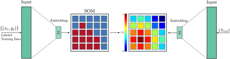
Prostate cancer (PCa) is the most frequent noncutaneous cancer in men. Early detection of PCa is essential for clinical decision making, and reducing metastasis and mortality rates. The current approach for PCa diagnosis is histopathologic analysis of core biopsies taken under transrectal ultrasound guidance (TRUS-guided). Both TRUS-guided systematic biopsy and MR-TRUS-guided fusion biopsy have limitations in accurately identifying PCa, intraoperatively. There is a need to augment this process by visualizing highly probable areas of PCa. Temporal enhanced ultrasound (TeUS) has emerged as a promising modality for PCa detection. Prior work focused on supervised classification of PCa verified by gold standard pathology. Pathology labels are noisy, and data from an entire core have a single label even when significantly heterogeneous. Additionally, supervised methods are limited by data from cores with known pathology, and a significant portion of prostate data is discarded without being used. We provide an end-to-end unsupervised solution to map PCa distribution from TeUS data using an innovative representation learning method, deep neural maps. TeUS data are transformed to a topologically arranged hyper-lattice, where similar samples are closer together in the lattice. Therefore, similar regions of malignant and benign tissue in the prostate are clustered together. Our proposed method increases the number of training samples by several orders of magnitude. Data from biopsy cores with known labels are used to associate the clusters with PCa. Cancer probability maps generated using the unsupervised clustering of TeUS data help intuitively visualize the distribution of abnormal tissue for augmenting TRUS-guided biopsies.
前列腺癌(PCa)是男性最常见的非皮肤癌。早期发现 PCa 对于临床决策制定、降低转移率和死亡率至关重要。目前,PCa 的诊断方法是在经直肠超声(TRUS)引导下进行核心活检的组织病理学分析。TRUS 引导的系统活检和 MR-TRUS 引导的融合活检在术中准确识别 PCa 方面都存在局限性。需要通过可视化高度可能存在 PCa 的区域来增强这一过程。时增强超声(TeUS)已成为一种很有前途的 PCa 检测方法。以前的工作主要集中在通过金标准病理学验证的 PCa 的监督分类上。病理学标签存在噪声,即使在明显异质的情况下,整个核心的数据也只有一个标签。此外,监督方法受到具有已知病理学的核心数据的限制,并且很大一部分前列腺数据未被使用就被丢弃了。我们使用创新的表示学习方法——深度神经图谱,为从 TeUS 数据映射 PCa 分布提供了一种端到端的无监督解决方案。TeUS 数据被转换为拓扑排列的超晶格,其中相似的样本在晶格中更接近。因此,前列腺中恶性和良性组织的相似区域被聚类在一起。我们提出的方法将训练样本的数量增加了几个数量级。使用具有已知标签的活检核心数据来关联聚类与 PCa。使用 TeUS 数据的无监督聚类生成的癌症概率图有助于直观地可视化异常组织的分布,从而增强 TRUS 引导的活检。