School of Chemical and Biomedical Engineering, Nanyang Technological University, Singapore, Singapore.
NTU-Northwestern Institute for Nanomedicine, Nanyang Technological University, Singapore, Singapore.
Nat Biomed Eng. 2018 Apr;2(4):227-238. doi: 10.1038/s41551-018-0218-x. Epub 2018 Apr 13.
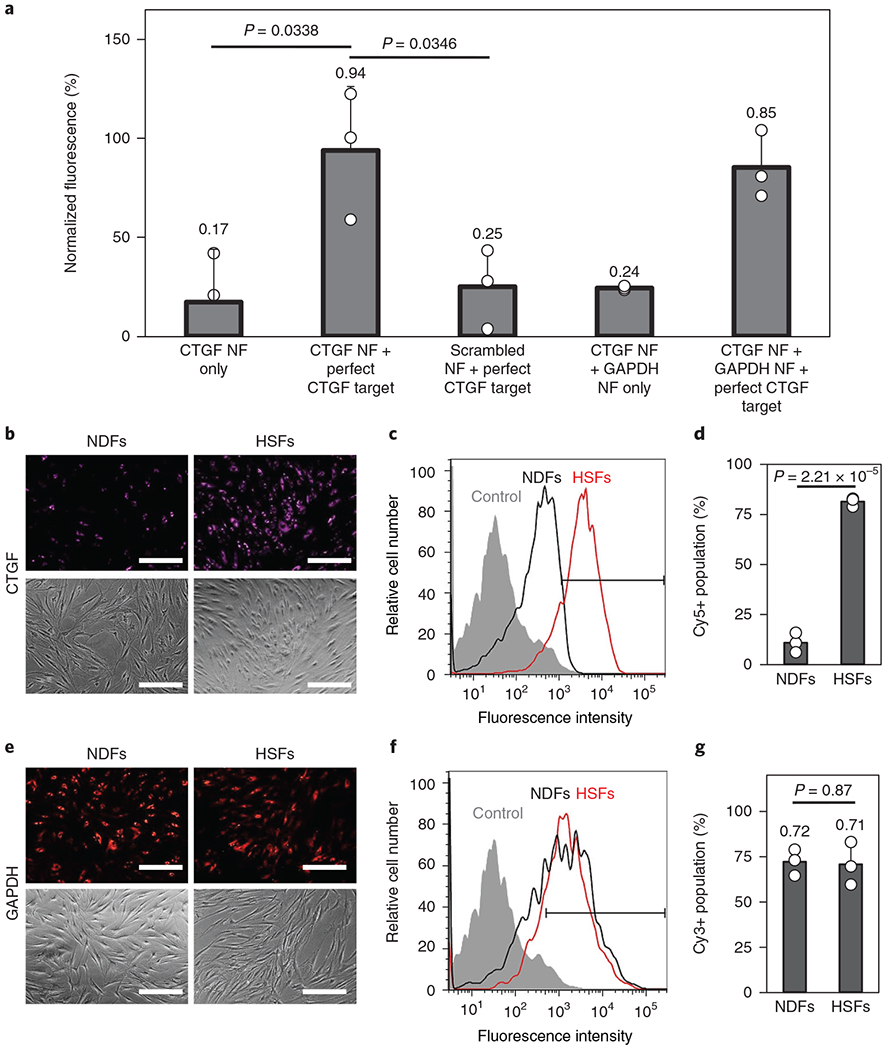
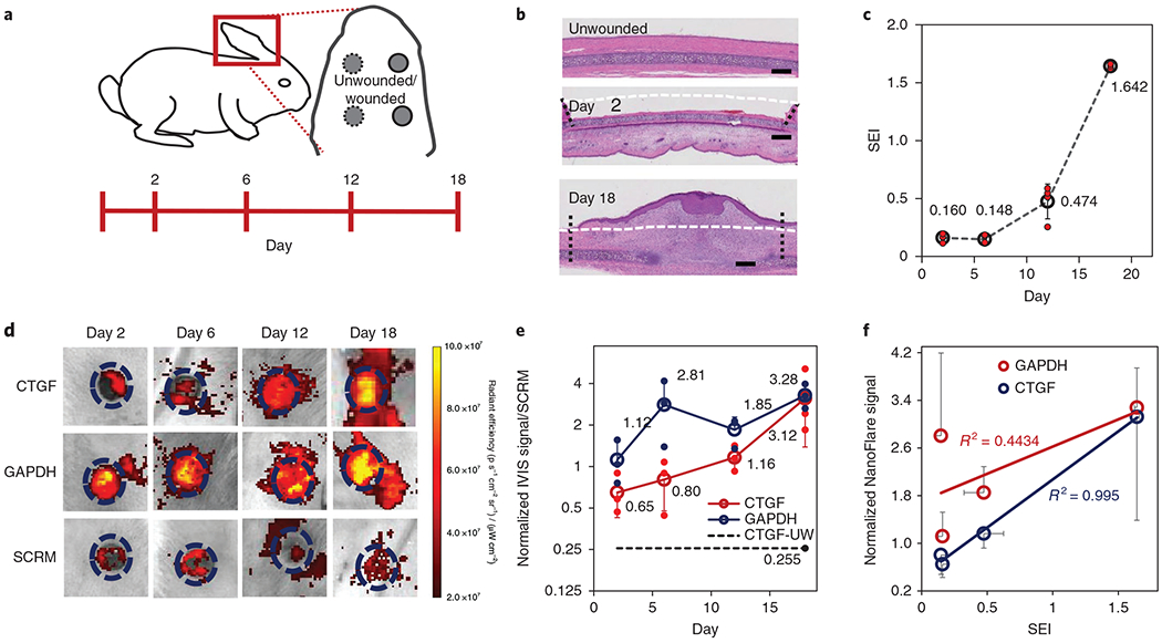
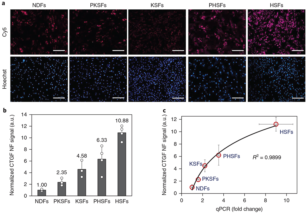
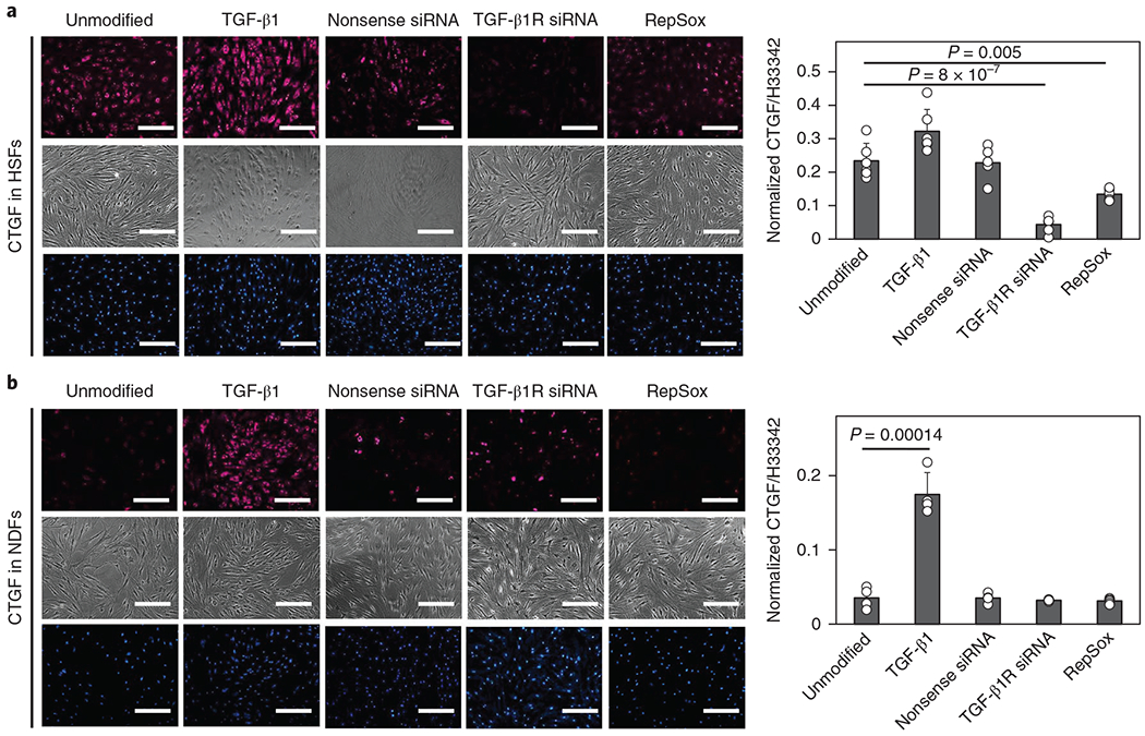
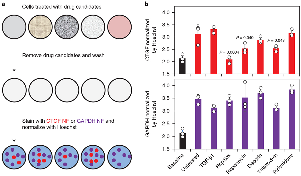
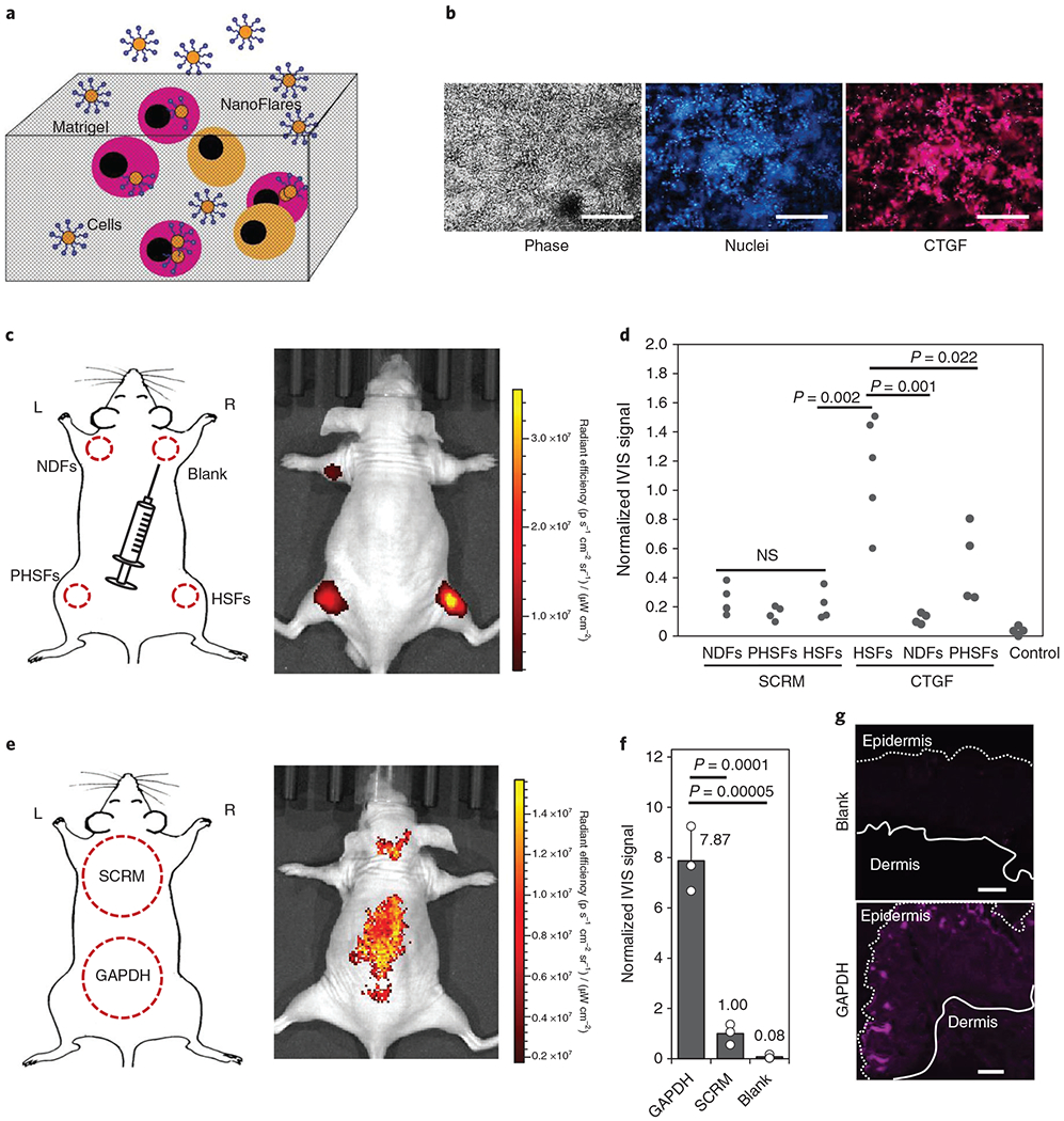
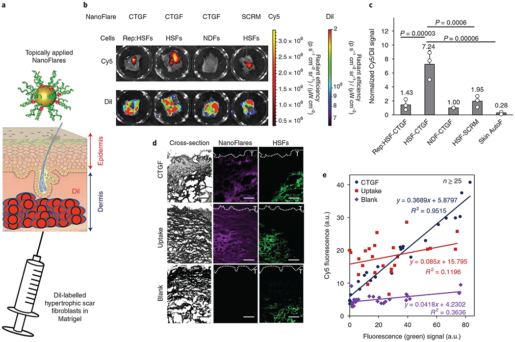
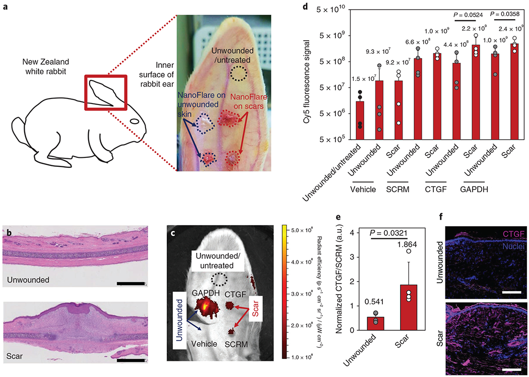
The accurate diagnosis of scar type and severity relies on histopathology of biopsied tissue, which is invasive and time-consuming, causes discomfort and may exacerbate scarring. Here, we show that imaging nanoprobes for the live-cell detection of intracellular messenger RNA (mRNA) (also known as NanoFlares) enable measurements of the expression of connective tissue growth factor (CTGF) as a visual indicator of hypertrophic scars and keloids. During cell culture, NanoFlares enabled the distinction of hypertrophic and keloidal fibroblasts from normal fibroblasts, and the detection of changes in CTGF expression resulting from the regulatory effects of transforming growth factor-β (TGF-β) agonists and TGF-β antagonists. We also applied the NanoFlares topically to the skin of live mice and rabbits, and to ex vivo human skin models. Transepidermal penetration of the NanoFlares enabled the visual and spectroscopic quantification of underlying abnormal fibroblasts on the basis of CTGF mRNA expression. Our proof-of-concept studies of topically applied NanoFlare technology as a means of biopsy-free scar diagnosis may eventually inform therapeutic decisions on the basis of the mRNA-expression patterns of skin disorders.
瘢痕类型和严重程度的准确诊断依赖于对活检组织进行组织病理学检查,这种方法具有侵袭性且耗时,会引起不适,并可能使瘢痕恶化。在这里,我们展示了用于活细胞内信使 RNA(mRNA)(也称为 NanoFlares)检测的成像纳米探针可用于测量结缔组织生长因子(CTGF)的表达,作为增生性瘢痕和瘢痕疙瘩的可视化指标。在细胞培养过程中,NanoFlares 能够区分增生性和成纤维细胞与正常成纤维细胞,并检测转化生长因子-β(TGF-β)激动剂和 TGF-β拮抗剂的调节作用导致的 CTGF 表达变化。我们还将 NanoFlares 局部应用于活小鼠和兔子的皮肤以及离体的人类皮肤模型。NanoFlares 的经表皮渗透使我们能够根据 CTGF mRNA 表达情况,对潜在异常成纤维细胞进行可视化和光谱定量。我们关于 NanoFlare 技术作为一种无活检瘢痕诊断方法的概念验证研究,最终可能会根据皮肤疾病的 mRNA 表达模式为治疗决策提供信息。