• 文献检索
  • 文档翻译
  • 深度研究
  • 学术资讯
  • Suppr Zotero 插件Zotero 插件
  • 邀请有礼
  • 套餐&价格
  • 历史记录
应用&插件
Suppr Zotero 插件Zotero 插件浏览器插件Mac 客户端Windows 客户端微信小程序
定价
高级版会员购买积分包购买API积分包
服务
文献检索文档翻译深度研究API 文档MCP 服务
关于我们
关于 Suppr公司介绍联系我们用户协议隐私条款
关注我们

Suppr 超能文献

核心技术专利:CN118964589B侵权必究
粤ICP备2023148730 号-1Suppr @ 2026

文献检索

告别复杂PubMed语法,用中文像聊天一样搜索,搜遍4000万医学文献。AI智能推荐,让科研检索更轻松。

立即免费搜索

文件翻译

保留排版,准确专业,支持PDF/Word/PPT等文件格式,支持 12+语言互译。

免费翻译文档

深度研究

AI帮你快速写综述,25分钟生成高质量综述,智能提取关键信息,辅助科研写作。

立即免费体验

基于不同空间分辨率的健康衰老和老年脑震荡计算机断层扫描的脑部分割

Brain Segmentation From Computed Tomography of Healthy Aging and Geriatric Concussion at Variable Spatial Resolutions.

作者信息

Irimia Andrei, Maher Alexander S, Rostowsky Kenneth A, Chowdhury Nahian F, Hwang Darryl H, Law E Meng

机构信息

USC Leonard Davis School of Gerontology, Ethel Percy Andrus Gerontology Center, University of Southern California, Los Angeles, CA, United States.

Department of Radiology, Keck School of Medicine of USC, University of Southern California, Los Angeles, CA, United States.

出版信息

Front Neuroinform. 2019 Mar 18;13:9. doi: 10.3389/fninf.2019.00009. eCollection 2019.

DOI:10.3389/fninf.2019.00009
PMID:30936828
原文链接:https://pmc.ncbi.nlm.nih.gov/articles/PMC6431646/
Abstract

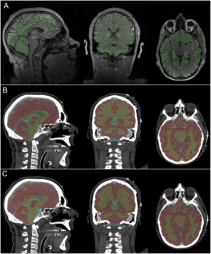
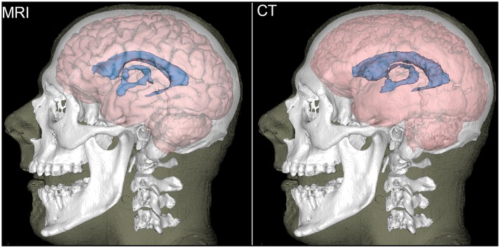
When properly implemented and processed, anatomic -weighted magnetic resonance imaging (MRI) can be ideal for the noninvasive quantification of white matter (WM) and gray matter (GM) in the living human brain. Although MRI is more suitable for distinguishing GM from WM than computed tomography (CT), the growing clinical use of the latter technique has renewed interest in head CT segmentation. Such interest is particularly strong in settings where MRI is unavailable, logistically unfeasible or prohibitively expensive. Nevertheless, whereas MRI segmentation is a sophisticated and technically-mature research field, the task of automatically classifying soft brain tissues from CT remains largely unexplored. Furthermore, brain segmentation methods for MRI hold considerable potential for adaptation and application to CT image processing. Here we demonstrate this by combining probabilistic, atlas-based classification with topologically-constrained tissue boundary refinement to delineate WM, GM and cerebrospinal fluid (CSF) from head CT images. The feasibility and utility of this approach are revealed by comparison of MRI-only vs. CT-only segmentations in geriatric concussion victims with both MRI and CT scans. Comparison of the two segmentations yields mean Sørensen-Dice coefficients of 85.5 ± 4.6% (WM), 86.7 ± 5.6% (GM) and 91.3 ± 2.8% (CSF), as well as average Hausdorff distances of 3.76 ± 1.85 mm (WM), 3.43 ± 1.53 mm (GM) and 2.46 ± 1.27 mm (CSF). Bootstrapping results suggest that the segmentation approach is sensitive enough to yield WM, GM and CSF volume estimates within ~5%, ~4%, and ~3% of their MRI-based estimates, respectively. To our knowledge, this is the first 3D segmentation approach for CT to undergo rigorous within-subject comparison with high-resolution MRI. Results suggest that (1) standard-quality CT allows WM/GM/CSF segmentation with reasonable accuracy, and that (2) the task of soft brain tissue classification from CT merits further attention from neuroimaging researchers.

摘要

当正确实施和处理时,解剖加权磁共振成像(MRI)对于活体人类大脑中白质(WM)和灰质(GM)的无创定量分析可能是理想的。尽管MRI比计算机断层扫描(CT)更适合区分GM和WM,但后一种技术在临床上的日益广泛应用重新引发了人们对头部CT分割的兴趣。在无法获得MRI、在后勤上不可行或成本过高的情况下,这种兴趣尤为强烈。然而,虽然MRI分割是一个复杂且技术成熟的研究领域,但从CT自动分类软脑组织的任务在很大程度上仍未得到探索。此外,MRI的脑部分割方法在适应和应用于CT图像处理方面具有相当大的潜力。在这里,我们通过将基于图谱的概率分类与拓扑约束的组织边界细化相结合,从头部CT图像中勾勒出WM、GM和脑脊液(CSF),从而证明了这一点。通过对同时进行MRI和CT扫描的老年脑震荡患者仅MRI分割与仅CT分割的比较,揭示了这种方法的可行性和实用性。两种分割的比较产生的平均索伦森 - 戴斯系数分别为85.5±4.6%(WM)、86.7±5.6%(GM)和91.3±2.8%(CSF),以及平均豪斯多夫距离分别为3.76±1.85毫米(WM)、3.43±1.53毫米(GM)和2.46±1.27毫米(CSF)。自展结果表明,分割方法足够灵敏,分别能产生WM、GM和CSF体积估计值,其与基于MRI的估计值的偏差分别在约5%、约4%和约3%以内。据我们所知,这是第一种用于CT的3D分割方法,可与高分辨率MRI进行严格的个体内比较。结果表明:(1)标准质量的CT能够以合理的准确性进行WM/GM/CSF分割;(2)从CT进行软脑组织分类的任务值得神经影像学研究人员进一步关注。

https://cdn.ncbi.nlm.nih.gov/pmc/blobs/e6bb/6431646/fbbbb027a0ee/fninf-13-00009-g0003.jpg
https://cdn.ncbi.nlm.nih.gov/pmc/blobs/e6bb/6431646/d69210423b76/fninf-13-00009-g0001.jpg
https://cdn.ncbi.nlm.nih.gov/pmc/blobs/e6bb/6431646/c0e3675d75ac/fninf-13-00009-g0002.jpg
https://cdn.ncbi.nlm.nih.gov/pmc/blobs/e6bb/6431646/fbbbb027a0ee/fninf-13-00009-g0003.jpg
https://cdn.ncbi.nlm.nih.gov/pmc/blobs/e6bb/6431646/d69210423b76/fninf-13-00009-g0001.jpg
https://cdn.ncbi.nlm.nih.gov/pmc/blobs/e6bb/6431646/c0e3675d75ac/fninf-13-00009-g0002.jpg
https://cdn.ncbi.nlm.nih.gov/pmc/blobs/e6bb/6431646/fbbbb027a0ee/fninf-13-00009-g0003.jpg

相似文献

1
Brain Segmentation From Computed Tomography of Healthy Aging and Geriatric Concussion at Variable Spatial Resolutions.基于不同空间分辨率的健康衰老和老年脑震荡计算机断层扫描的脑部分割
Front Neuroinform. 2019 Mar 18;13:9. doi: 10.3389/fninf.2019.00009. eCollection 2019.
2
A reliable spatially normalized template of the human spinal cord--Applications to automated white matter/gray matter segmentation and tensor-based morphometry (TBM) mapping of gray matter alterations occurring with age.一个可靠的人类脊髓空间标准化模板——应用于自动白质/灰质分割以及基于张量的形态测量学(TBM)对与年龄相关的灰质变化进行映射。
Neuroimage. 2015 Aug 15;117:20-8. doi: 10.1016/j.neuroimage.2015.05.034. Epub 2015 May 21.
3
Automatic segmentation of MR brain images of preterm infants using supervised classification.使用监督分类对早产儿脑部磁共振图像进行自动分割。
Neuroimage. 2015 Sep;118:628-41. doi: 10.1016/j.neuroimage.2015.06.007. Epub 2015 Jun 7.
4
Decomposing the Hounsfield unit: probabilistic segmentation of brain tissue in computed tomography.分解 Hounsfield 单位:计算机断层扫描中脑组织的概率分割。
Clin Neuroradiol. 2012 Mar;22(1):79-91. doi: 10.1007/s00062-011-0123-0. Epub 2012 Jan 21.
5
A discriminative model-constrained EM approach to 3D MRI brain tissue classification and intensity non-uniformity correction.基于判别模型约束的 EM 方法进行 3D MRI 脑组织结构分类和强度不均匀性校正。
Phys Med Biol. 2011 Jun 7;56(11):3269-300. doi: 10.1088/0031-9155/56/11/007. Epub 2011 May 10.
6
Learning-based 3T brain MRI segmentation with guidance from 7T MRI labeling.基于学习的3T脑磁共振成像分割,由7T磁共振成像标记引导。
Med Phys. 2016 Dec;43(12):6588-6597. doi: 10.1118/1.4967487.
7
Deep learning from MRI-derived labels enables automatic brain tissue classification on human brain CT.基于 MRI 标记的深度学习可实现人脑 CT 上的自动脑组织分类。
Neuroimage. 2021 Dec 1;244:118606. doi: 10.1016/j.neuroimage.2021.118606. Epub 2021 Sep 25.
8
Automatic segmentation of cerebrospinal fluid, white and gray matter in unenhanced computed tomography images.脑实质、脑白质和脑脊髓液在未增强 CT 图像中的自动分割。
Acad Radiol. 2010 Nov;17(11):1350-8. doi: 10.1016/j.acra.2010.06.005. Epub 2010 Jul 15.
9
White Matter and Gray Matter Segmentation in 4D Computed Tomography.四维计算机断层扫描中的脑白质和脑灰质分割。
Sci Rep. 2017 Mar 9;7(1):119. doi: 10.1038/s41598-017-00239-z.
10
Segmentation of gray matter, white matter, and CSF with fluid and white matter suppression using MP2RAGE.采用 MP2RAGE 对灰质、白质和 CSF 进行分割,同时抑制脑脊液和白质信号。
J Magn Reson Imaging. 2018 Dec;48(6):1540-1550. doi: 10.1002/jmri.26014. Epub 2018 Mar 22.

引用本文的文献

1
Physical activity mediates age differences in cognition among Tsimane forager-horticulturalists.体力活动介导了齐曼内觅食-园艺人群认知能力的年龄差异。
J Gerontol A Biol Sci Med Sci. 2025 Jul 24;80(8). doi: 10.1093/gerona/glaf163.
2
Refining CT image analysis: Exploring adaptive fusion in U-nets for enhanced brain tissue segmentation.优化CT图像分析:探索U-Net中的自适应融合以增强脑组织分割
PLoS One. 2025 Jun 11;20(6):e0323692. doi: 10.1371/journal.pone.0323692. eCollection 2025.
3
Rapidly progressive brain atrophy in ventilated patients: a retrospective descriptive study.

本文引用的文献

1
Optimization and comparative evaluation of nonlinear deformation algorithms for atlas-based segmentation of DBS target nuclei.基于图谱的 DBS 靶点分割的非线性变形算法的优化与比较评价。
Neuroimage. 2019 Jan 1;184:586-598. doi: 10.1016/j.neuroimage.2018.09.061. Epub 2018 Sep 26.
2
Imaging in Neurocritical Care Practice.神经危重症护理实践中的影像学。
Semin Respir Crit Care Med. 2017 Dec;38(6):840-852. doi: 10.1055/s-0037-1608770. Epub 2017 Dec 20.
3
Reproducibility of Brain MRI Segmentation Algorithms: Empirical Comparison of Local MAP PSTAPLE, FreeSurfer, and FSL-FIRST.
机械通气患者的快速进行性脑萎缩:一项回顾性描述性研究。
Sci Rep. 2024 Nov 29;14(1):29729. doi: 10.1038/s41598-024-81372-4.
4
BounTI (boundary-preserving threshold iteration): A user-friendly tool for automatic hard tissue segmentation.BounTI(边界保持阈值迭代):一个用于自动硬组织分割的用户友好工具。
J Anat. 2024 Dec;245(6):829-841. doi: 10.1111/joa.14063. Epub 2024 May 17.
5
Increases in regional brain volume across two native South American male populations.两个南美男性群体的区域性脑容量增加。
Geroscience. 2024 Oct;46(5):4563-4583. doi: 10.1007/s11357-024-01168-2. Epub 2024 Apr 29.
6
Poor Oral Health Is Associated With Inflammation, Aortic Valve Calcification, and Brain Volume Among Forager-Farmers.口腔健康状况不佳与炎症、主动脉瓣钙化和觅食-农耕者的大脑体积有关。
J Gerontol A Biol Sci Med Sci. 2024 May 1;79(5). doi: 10.1093/gerona/glae013.
7
Rapidly progressive cerebral atrophy following a posterior cranial fossa stroke: Assessment with semiautomatic CT volumetry.颅后窝卒中后迅速进展性脑萎缩:半自动化 CT 容积测量评估。
Acta Neurochir (Wien). 2023 Jun;165(6):1575-1584. doi: 10.1007/s00701-023-05609-3. Epub 2023 Apr 29.
8
Mapping spreading depolarisations after traumatic brain injury: a pilot clinical study protocol.创伤性脑损伤后扩散去极化的定位:一项初步临床研究方案。
BMJ Open. 2022 Jul 13;12(7):e061663. doi: 10.1136/bmjopen-2022-061663.
9
Deep learning-based automated segmentation of eight brain anatomical regions using head CT images in PET/CT.基于深度学习的头部 CT 图像在正电子发射断层扫描/计算机断层扫描中对 8 个脑解剖区域的自动分割。
BMC Med Imaging. 2022 May 26;22(1):99. doi: 10.1186/s12880-022-00807-4.
10
A Comparison of Global Brain Volumetrics Obtained from CT versus MRI Using 2 Publicly Available Software Packages.使用 2 个公开可用的软件包比较 CT 与 MRI 获得的全球脑容量。
AJNR Am J Neuroradiol. 2022 Feb;43(2):245-250. doi: 10.3174/ajnr.A7403.
脑磁共振成像分割算法的可重复性:局部MAP PSTAPLE、FreeSurfer和FSL-FIRST的实证比较
J Neuroimaging. 2018 Mar;28(2):162-172. doi: 10.1111/jon.12483. Epub 2017 Nov 14.
4
Coronary atherosclerosis in indigenous South American Tsimane: a cross-sectional cohort study.南美原住民提斯曼人中的冠状动脉粥样硬化:一项横断面队列研究。
Lancet. 2017 Apr 29;389(10080):1730-1739. doi: 10.1016/S0140-6736(17)30752-3. Epub 2017 Mar 17.
5
White Matter and Gray Matter Segmentation in 4D Computed Tomography.四维计算机断层扫描中的脑白质和脑灰质分割。
Sci Rep. 2017 Mar 9;7(1):119. doi: 10.1038/s41598-017-00239-z.
6
Enhanced gray-white matter differentiation on non-enhanced CT using a frequency selective non-linear blending.使用频率选择性非线性融合技术在非增强CT上增强灰白质区分度。
Neuroradiology. 2016 Jul;58(7):649-55. doi: 10.1007/s00234-016-1674-1. Epub 2016 Mar 10.
7
Test-retest reliability of freesurfer measurements within and between sites: Effects of visual approval process.Freesurfer测量在不同站点内部和之间的重测信度:视觉审批流程的影响。
Hum Brain Mapp. 2015 Sep;36(9):3472-85. doi: 10.1002/hbm.22856. Epub 2015 May 28.
8
Improvement of image quality and radiation dose of CT perfusion of the brain by means of low-tube voltage (70 KV).通过低管电压(70千伏)改善脑部CT灌注成像的图像质量和辐射剂量。
Eur Radiol. 2014 Aug;24(8):1906-13. doi: 10.1007/s00330-014-3247-1. Epub 2014 Jun 4.
9
Recent and future directions in CT imaging.CT成像的近期及未来发展方向。
Ann Biomed Eng. 2014 Feb;42(2):260-8. doi: 10.1007/s10439-014-0974-z. Epub 2014 Jan 17.
10
Quantifying the reliability of image replication studies: the image intraclass correlation coefficient (I2C2).量化图像复制研究的可靠性:图像组内相关系数(I2C2)。
Cogn Affect Behav Neurosci. 2013 Dec;13(4):714-24. doi: 10.3758/s13415-013-0196-0.