• 文献检索
  • 文档翻译
  • 深度研究
  • 学术资讯
  • Suppr Zotero 插件Zotero 插件
  • 邀请有礼
  • 套餐&价格
  • 历史记录
应用&插件
Suppr Zotero 插件Zotero 插件浏览器插件Mac 客户端Windows 客户端微信小程序
定价
高级版会员购买积分包购买API积分包
服务
文献检索文档翻译深度研究API 文档MCP 服务
关于我们
关于 Suppr公司介绍联系我们用户协议隐私条款
关注我们

Suppr 超能文献

核心技术专利:CN118964589B侵权必究
粤ICP备2023148730 号-1Suppr @ 2026

文献检索

告别复杂PubMed语法,用中文像聊天一样搜索,搜遍4000万医学文献。AI智能推荐,让科研检索更轻松。

立即免费搜索

文件翻译

保留排版,准确专业,支持PDF/Word/PPT等文件格式,支持 12+语言互译。

免费翻译文档

深度研究

AI帮你快速写综述,25分钟生成高质量综述,智能提取关键信息,辅助科研写作。

立即免费体验

全切片成像中的数字图像分析介绍:数字病理协会白皮书

Introduction to Digital Image Analysis in Whole-slide Imaging: A White Paper from the Digital Pathology Association.

作者信息

Aeffner Famke, Zarella Mark D, Buchbinder Nathan, Bui Marilyn M, Goodman Matthew R, Hartman Douglas J, Lujan Giovanni M, Molani Mariam A, Parwani Anil V, Lillard Kate, Turner Oliver C, Vemuri Venkata N P, Yuil-Valdes Ana G, Bowman Douglas

机构信息

Amgen Inc., Amgen Research, Comparative Biology and Safety Sciences, South San Francisco, CA, USA.

Department of Pathology and Laboratory Medicine, Drexel University, College of Medicine, Philadelphia, PA, USA.

出版信息

J Pathol Inform. 2019 Mar 8;10:9. doi: 10.4103/jpi.jpi_82_18. eCollection 2019.

DOI:10.4103/jpi.jpi_82_18
PMID:30984469
原文链接:https://pmc.ncbi.nlm.nih.gov/articles/PMC6437786/
Abstract

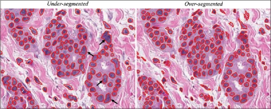
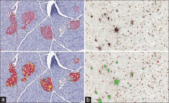
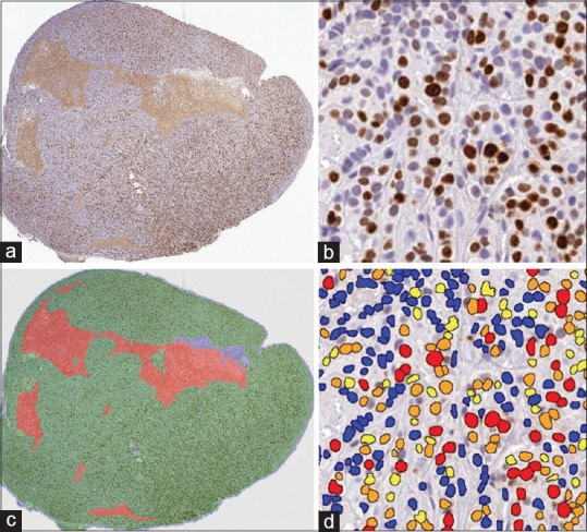
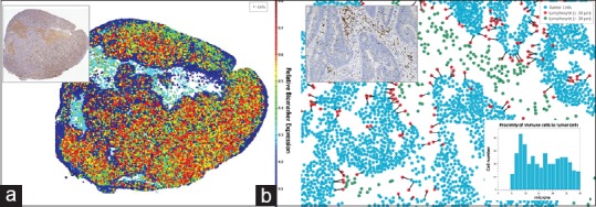
The advent of whole-slide imaging in digital pathology has brought about the advancement of computer-aided examination of tissue via digital image analysis. Digitized slides can now be easily annotated and analyzed via a variety of algorithms. This study reviews the fundamentals of tissue image analysis and aims to provide pathologists with basic information regarding the features, applications, and general workflow of these new tools. The review gives an overview of the basic categories of software solutions available, potential analysis strategies, technical considerations, and general algorithm readouts. Advantages and limitations of tissue image analysis are discussed, and emerging concepts, such as artificial intelligence and machine learning, are introduced. Finally, examples of how digital image analysis tools are currently being used in diagnostic laboratories, translational research, and drug development are discussed.

摘要

数字病理学中全切片成像的出现推动了通过数字图像分析对组织进行计算机辅助检查的发展。现在,数字化切片可以通过各种算法轻松进行标注和分析。本研究回顾了组织图像分析的基本原理,旨在为病理学家提供有关这些新工具的特征、应用和一般工作流程的基本信息。该综述概述了可用软件解决方案的基本类别、潜在分析策略、技术考量和一般算法输出。讨论了组织图像分析的优点和局限性,并介绍了人工智能和机器学习等新兴概念。最后讨论了数字图像分析工具目前在诊断实验室、转化研究和药物开发中的应用实例。

https://cdn.ncbi.nlm.nih.gov/pmc/blobs/e0c0/6437786/701e0caf269d/JPI-10-9-g005.jpg
https://cdn.ncbi.nlm.nih.gov/pmc/blobs/e0c0/6437786/5264c209a7fb/JPI-10-9-g001.jpg
https://cdn.ncbi.nlm.nih.gov/pmc/blobs/e0c0/6437786/755d01b42602/JPI-10-9-g002.jpg
https://cdn.ncbi.nlm.nih.gov/pmc/blobs/e0c0/6437786/b20b935b8c9a/JPI-10-9-g003.jpg
https://cdn.ncbi.nlm.nih.gov/pmc/blobs/e0c0/6437786/4f257d049093/JPI-10-9-g004.jpg
https://cdn.ncbi.nlm.nih.gov/pmc/blobs/e0c0/6437786/701e0caf269d/JPI-10-9-g005.jpg

相似文献

1
Introduction to Digital Image Analysis in Whole-slide Imaging: A White Paper from the Digital Pathology Association.全切片成像中的数字图像分析介绍:数字病理协会白皮书
J Pathol Inform. 2019 Mar 8;10:9. doi: 10.4103/jpi.jpi_82_18. eCollection 2019.
2
Whole-slide imaging, tissue image analysis, and artificial intelligence in veterinary pathology: An updated introduction and review.全玻片成像、组织图像分析和兽医病理学中的人工智能:更新介绍和综述。
Vet Pathol. 2022 Jan;59(1):6-25. doi: 10.1177/03009858211040484. Epub 2021 Sep 14.
3
Digital pathology and artificial intelligence.数字病理学与人工智能。
Lancet Oncol. 2019 May;20(5):e253-e261. doi: 10.1016/S1470-2045(19)30154-8.
4
Digital Microscopy, Image Analysis, and Virtual Slide Repository.数字显微镜、图像分析与虚拟切片库
ILAR J. 2018 Dec 1;59(1):66-79. doi: 10.1093/ilar/ily007.
5
Special Issue on Digital Pathology, Tissue Image Analysis, Artificial Intelligence, and Machine Learning: Approximation of the Effect of Novel Technologies on Toxicologic Pathology.数字病理学、组织图像分析、人工智能和机器学习特刊:新型技术对毒理学病理学影响的近似。
Toxicol Pathol. 2021 Jun;49(4):705-708. doi: 10.1177/0192623321993756. Epub 2021 Apr 12.
6
Applications of Digital and Computational Pathology and Artificial Intelligence in Genitourinary Pathology Diagnostics.数字和计算病理学与人工智能在泌尿生殖系统病理学诊断中的应用。
Surg Pathol Clin. 2022 Dec;15(4):759-785. doi: 10.1016/j.path.2022.08.001.
7
Whole Slide Images in Artificial Intelligence Applications in Digital Pathology: Challenges and Pitfalls.人工智能在数字病理学中的全切片图像应用:挑战与陷阱。
Turk Patoloji Derg. 2023;39(2):101-108. doi: 10.5146/tjpath.2023.01601.
8
Biobanking in the digital pathology era.数字病理学时代的生物样本库。
Oncol Res. 2022 Aug 31;29(4):229-233. doi: 10.32604/or.2022.024892. eCollection 2021.
9
Image analysis and machine learning in digital pathology: Challenges and opportunities.数字病理学中的图像分析与机器学习:挑战与机遇
Med Image Anal. 2016 Oct;33:170-175. doi: 10.1016/j.media.2016.06.037. Epub 2016 Jul 4.
10
Digital pathology and artificial intelligence as the next chapter in diagnostic hematopathology.数字病理学与人工智能作为诊断血液病理学的新篇章。
Semin Diagn Pathol. 2023 Mar;40(2):88-94. doi: 10.1053/j.semdp.2023.02.001. Epub 2023 Feb 15.

引用本文的文献

1
Innovative approach for the qualitative-quantitative assessment of neurodevelopment biomarkers research in placenta tissue using immunohistochemistry digital image analysis.使用免疫组织化学数字图像分析对胎盘组织中神经发育生物标志物研究进行定性-定量评估的创新方法。
Biol Methods Protoc. 2025 Jul 11;10(1):bpaf056. doi: 10.1093/biomethods/bpaf056. eCollection 2025.
2
An open-source platform for structured annotation and computational workflows in digital pathology research.一个用于数字病理学研究中结构化注释和计算工作流程的开源平台。
Sci Rep. 2025 Aug 7;15(1):28910. doi: 10.1038/s41598-025-13546-7.
3
Automating Colon Polyp Classification in Digital Pathology by Evaluation of a "Machine Learning as a Service" AI Model: Algorithm Development and Validation Study.

本文引用的文献

1
From Detection of Individual Metastases to Classification of Lymph Node Status at the Patient Level: The CAMELYON17 Challenge.从检测单个转移灶到对患者水平的淋巴结状态进行分类:CAMELYON17 挑战赛。
IEEE Trans Med Imaging. 2019 Feb;38(2):550-560. doi: 10.1109/TMI.2018.2867350.
2
Quantitative Image Analysis of Human Epidermal Growth Factor Receptor 2 Immunohistochemistry for Breast Cancer: Guideline From the College of American Pathologists.人类表皮生长因子受体 2 免疫组织化学乳腺癌的定量图像分析:美国病理学家学院指南。
Arch Pathol Lab Med. 2019 Oct;143(10):1180-1195. doi: 10.5858/arpa.2018-0378-CP. Epub 2019 Jan 15.
3
通过评估“机器学习即服务”人工智能模型实现数字病理学中结肠息肉分类的自动化:算法开发与验证研究
JMIR Form Res. 2025 Jul 31;9:e67457. doi: 10.2196/67457.
4
Comparing non-machine learning vs. machine learning methods for Ki67 scoring in gastrointestinal neuroendocrine tumors.比较非机器学习方法与机器学习方法在胃肠道神经内分泌肿瘤中进行Ki67评分的情况。
Sci Rep. 2025 Jul 29;15(1):27700. doi: 10.1038/s41598-025-08778-6.
5
Segmentation and quantification of testicular histology images using machine learning bioimage analysis tools; Ilastik and Fiji software.使用机器学习生物图像分析工具(Ilastik和Fiji软件)对睾丸组织学图像进行分割和量化
MethodsX. 2025 Jul 11;15:103503. doi: 10.1016/j.mex.2025.103503. eCollection 2025 Dec.
6
Evaluation of radiation-induced bone marrow toxicity using artificial intelligence-based image analysis in mice.利用基于人工智能的图像分析评估小鼠辐射诱导的骨髓毒性。
Toxicol Res. 2025 Apr 8;41(4):397-405. doi: 10.1007/s43188-025-00290-3. eCollection 2025 Jul.
7
Rapid 3D imaging at cellular resolution for digital cytopathology with a multi-camera array scanner (MCAS).使用多相机阵列扫描仪(MCAS)以细胞分辨率进行快速3D成像用于数字细胞病理学。
Npj Imaging. 2024 Oct 1;2(1):39. doi: 10.1038/s44303-024-00042-2.
8
Integrated histopathology of the human pancreas throughout stages of type 1 diabetes progression.1型糖尿病进展各阶段人类胰腺的综合组织病理学研究。
Res Sq. 2025 Jun 10:rs.3.rs-6673858. doi: 10.21203/rs.3.rs-6673858/v1.
9
Artificial intelligence in cancer pathology: Applications, challenges, and future directions.癌症病理学中的人工智能:应用、挑战及未来方向。
Cytojournal. 2025 Apr 19;22:45. doi: 10.25259/Cytojournal_272_2024. eCollection 2025.
10
Comparison of the efficiency of digital pathology with the conventional methodology for the diagnosis of biopsies in an anatomical pathology laboratory in Spain.西班牙一家解剖病理学实验室中数字病理学与传统方法在活检诊断效率方面的比较。
J Pathol Inform. 2025 Apr 1;17:100439. doi: 10.1016/j.jpi.2025.100439. eCollection 2025 Apr.
BCL-2 expression aids in the immunohistochemical prediction of the Oncotype DX breast cancer recurrence score.
BCL-2表达有助于Oncotype DX乳腺癌复发评分的免疫组织化学预测。
BMC Clin Pathol. 2018 Dec 18;18:14. doi: 10.1186/s12907-018-0082-3. eCollection 2018.
4
Digital Microscopy, Image Analysis, and Virtual Slide Repository.数字显微镜、图像分析与虚拟切片库
ILAR J. 2018 Dec 1;59(1):66-79. doi: 10.1093/ilar/ily007.
5
A Practical Guide to Whole Slide Imaging: A White Paper From the Digital Pathology Association.全切片成像实用指南:数字病理学协会白皮书。
Arch Pathol Lab Med. 2019 Feb;143(2):222-234. doi: 10.5858/arpa.2018-0343-RA. Epub 2018 Oct 11.
6
Machine Learning Methods for Histopathological Image Analysis.用于组织病理学图像分析的机器学习方法
Comput Struct Biotechnol J. 2018 Feb 9;16:34-42. doi: 10.1016/j.csbj.2018.01.001. eCollection 2018.
7
Validation of a Muscle-Specific Tissue Image Analysis Tool for Quantitative Assessment of Dystrophin Staining in Frozen Muscle Biopsies.肌肉特异性组织图像分析工具定量评估冰冻肌肉活检中肌营养不良蛋白染色的验证。
Arch Pathol Lab Med. 2019 Feb;143(2):197-205. doi: 10.5858/arpa.2017-0536-OA. Epub 2018 Aug 31.
8
eDoctor: machine learning and the future of medicine.医生:机器学习与医学的未来。
J Intern Med. 2018 Dec;284(6):603-619. doi: 10.1111/joim.12822. Epub 2018 Sep 3.
9
Quanti.us: a tool for rapid, flexible, crowd-based annotation of images.Quanti.us:一种用于快速、灵活、基于众包的图像标注工具。
Nat Methods. 2018 Aug;15(8):587-590. doi: 10.1038/s41592-018-0069-0. Epub 2018 Jul 31.
10
Nuclear shape and orientation features from H&E images predict survival in early-stage estrogen receptor-positive breast cancers.H&E 图像的核形状和定向特征可预测早期雌激素受体阳性乳腺癌的生存情况。
Lab Invest. 2018 Nov;98(11):1438-1448. doi: 10.1038/s41374-018-0095-7. Epub 2018 Jun 29.