• 文献检索
  • 文档翻译
  • 深度研究
  • 学术资讯
  • Suppr Zotero 插件Zotero 插件
  • 邀请有礼
  • 套餐&价格
  • 历史记录
应用&插件
Suppr Zotero 插件Zotero 插件浏览器插件Mac 客户端Windows 客户端微信小程序
定价
高级版会员购买积分包购买API积分包
服务
文献检索文档翻译深度研究API 文档MCP 服务
关于我们
关于 Suppr公司介绍联系我们用户协议隐私条款
关注我们

Suppr 超能文献

核心技术专利:CN118964589B侵权必究
粤ICP备2023148730 号-1Suppr @ 2026

文献检索

告别复杂PubMed语法,用中文像聊天一样搜索,搜遍4000万医学文献。AI智能推荐,让科研检索更轻松。

立即免费搜索

文件翻译

保留排版,准确专业,支持PDF/Word/PPT等文件格式,支持 12+语言互译。

免费翻译文档

深度研究

AI帮你快速写综述,25分钟生成高质量综述,智能提取关键信息,辅助科研写作。

立即免费体验

内镜黏膜下剥离术中使用双红光成像的止血治疗:一项多中心、开放标签、随机对照试验。

Haemostasis treatment using dual red imaging during endoscopic submucosal dissection: a multicentre, open-label, randomised controlled trial.

作者信息

Fujimoto Ai, Saito Yutaka, Abe Seiirhicro, Hoteya Shu, Nomura Kosuke, Yasuda Hiroshi, Matsuo Yasumasa, Uraoka Toshio, Kuribayashi Shiko, Saito Itaru, Tsuji Yosuke, Maehata Tadateru, Ochiai Yasutoshi, Nishizawa Toshihiro, Yahagi Naohisa

机构信息

Division of Research and Development for Minimally Invasive Treatment, Cancer Center, Keio University School of Medicine, Tokyo, Japan.

Department of Gastroenterology and Hepatology, National Hospital Organization Tokyo Medical Center, Tokyo, Japan.

出版信息

BMJ Open Gastroenterol. 2019 Mar 30;6(1):e000275. doi: 10.1136/bmjgast-2019-000275. eCollection 2019.

DOI:10.1136/bmjgast-2019-000275
PMID:30997140
原文链接:https://pmc.ncbi.nlm.nih.gov/articles/PMC6441255/
Abstract

INTRODUCTION

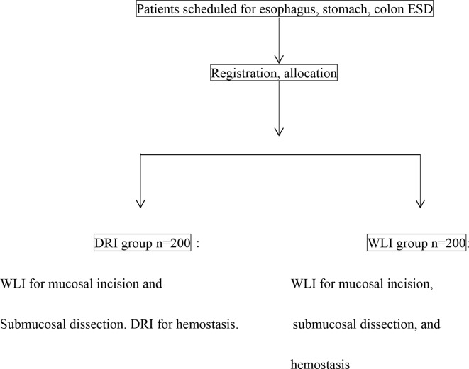
Patients scheduled to undergo oesophageal, gastric and colorectal endoscopic submucosal dissection (ESD) are to be investigated to verify the efficacy of dual red imaging (DRI) for establishing haemostasis during ESD.

METHODS AND ANALYSIS

The trial is designed as a multicentre, open-label randomised, parallel-group, controlled intervention study. Registered patients will be randomly assigned to DRI and white light imaging (WLI) groups. In the DRI group, the mucosal incision and submucosal dissection will be performed by WLI, and haemostasis will be managed by DRI when bleeding occurs. In the WLI group, the mucosal incision and submucosal dissection are to be performed by WLI and the haemostasis management is to be performed by WLI. The primary endpoint is the time from the recognition of bleeding up to the achievement of complete haemostasis (haemostasis time). The secondary endpoints are the operation time, the proportion of cases in which perforation occurs, and the psychological stress experienced by the endoscopist during haemostasis treatment.

ETHICS AND DISSEMINATION

This trial was approved by the Keio University Review Board for Clinical Trials (5 December 2016).

DISCUSSION

This will be the first multicentre collaborative research using DRI for haemostasis treatment during ESD. When the safety and simplicity of DRI as a treatment for haemostasis during ESD can be proven, the ESD procedure can be simplified and disseminated more widely in clinical practice.

TRIAL REGISTRATION NUMBER

UMIN000025134.

摘要

引言

计划接受食管、胃和结肠内镜黏膜下剥离术(ESD)的患者将接受调查,以验证双波长成像(DRI)在ESD期间建立止血效果方面的作用。

方法与分析

该试验设计为一项多中心、开放标签、随机、平行组对照干预研究。登记的患者将被随机分配至DRI组和白光成像(WLI)组。在DRI组中,黏膜切开和黏膜下剥离将通过WLI进行,出血时通过DRI进行止血处理。在WLI组中,黏膜切开和黏膜下剥离通过WLI进行,止血处理也通过WLI进行。主要终点是从识别出血到实现完全止血的时间(止血时间)。次要终点是手术时间、穿孔发生病例的比例以及内镜医师在止血治疗期间所经历的心理压力。

伦理与传播

本试验已获得庆应义塾大学临床试验审查委员会批准(2016年12月5日)。

讨论

这将是第一项使用DRI进行ESD期间止血治疗的多中心合作研究。当DRI作为ESD期间止血治疗的安全性和简便性得到证实时,ESD操作可以简化并在临床实践中更广泛地推广。

试验注册号

UMIN000025134。

https://cdn.ncbi.nlm.nih.gov/pmc/blobs/ab3c/6441255/15c458a7b211/bmjgast-2019-000275f04.jpg
https://cdn.ncbi.nlm.nih.gov/pmc/blobs/ab3c/6441255/cb25a0d38934/bmjgast-2019-000275f01.jpg
https://cdn.ncbi.nlm.nih.gov/pmc/blobs/ab3c/6441255/51dce609e708/bmjgast-2019-000275f02.jpg
https://cdn.ncbi.nlm.nih.gov/pmc/blobs/ab3c/6441255/11609c4aac5f/bmjgast-2019-000275f03.jpg
https://cdn.ncbi.nlm.nih.gov/pmc/blobs/ab3c/6441255/15c458a7b211/bmjgast-2019-000275f04.jpg

相似文献

1
Haemostasis treatment using dual red imaging during endoscopic submucosal dissection: a multicentre, open-label, randomised controlled trial.内镜黏膜下剥离术中使用双红光成像的止血治疗:一项多中心、开放标签、随机对照试验。
BMJ Open Gastroenterol. 2019 Mar 30;6(1):e000275. doi: 10.1136/bmjgast-2019-000275. eCollection 2019.
2
Clinical usefulness of red dichromatic imaging in hemostatic treatment during endoscopic submucosal dissection: First report from a multicenter, open-label, randomized controlled trial.内镜黏膜下剥离术中红色双折射成像在止血治疗中的临床应用:多中心、开放标签、随机对照试验的首次报告。
Dig Endosc. 2022 Jan;34(2):379-390. doi: 10.1111/den.14191. Epub 2021 Dec 20.
3
Efficacy of using red dichromatic imaging throughout endoscopic submucosal dissection procedure.在整个内镜黏膜下剥离手术过程中使用红色双色成像的疗效。
Surg Endosc. 2023 Jan;37(1):503-509. doi: 10.1007/s00464-022-09543-w. Epub 2022 Aug 24.
4
Efficacy of a new image-enhancement technique for achieving hemostasis in endoscopic submucosal dissection.一种新的图像增强技术在实现内镜黏膜下剥离术止血中的疗效。
Gastrointest Endosc. 2020 Sep;92(3):667-674. doi: 10.1016/j.gie.2020.05.033. Epub 2020 Jun 1.
5
Comparison of the procedure time differences between hybrid endoscopic submucosal dissection and conventional endoscopic submucosal dissection in patients with early gastric neoplasms: a study protocol for a multi-center randomized controlled trial (Hybrid-G trial).比较早期胃癌患者行内镜黏膜下剥离术与杂交内镜黏膜下剥离术的手术时间差异:一项多中心随机对照试验研究方案(Hybrid-G 试验)
Trials. 2022 Feb 21;23(1):166. doi: 10.1186/s13063-022-06099-x.
6
Clinical Usefulness of Dual Red Imaging in Gastric Endoscopic Submucosal Dissection: A Pilot Study.双红成像在胃内镜黏膜下剥离术中的临床应用:一项初步研究。
Clin Endosc. 2020 Jan;53(1):54-59. doi: 10.5946/ce.2019.065. Epub 2019 Sep 3.
7
Dual Red Imaging Maintains Clear Visibility During Colorectal Endoscopic Submucosal Dissection.双红成像在结直肠内镜黏膜下剥离术中保持清晰可视性。
Dig Dis Sci. 2019 Jan;64(1):224-231. doi: 10.1007/s10620-018-5306-y. Epub 2018 Oct 4.
8
Clinical impact of dual red imaging in colorectal endoscopic submucosal dissection: a pilot study.双红成像在大肠内镜黏膜下剥离术中的临床应用:一项前瞻性研究
Therap Adv Gastroenterol. 2016 Sep;9(5):679-83. doi: 10.1177/1756283X16645501. Epub 2016 May 4.
9
Red Dichromatic Imaging Improves the Recognition of Bleeding Points During Endoscopic Submucosal Dissection.红色双色色差成像提高内镜黏膜下剥离术中出血点的识别率。
Dig Dis Sci. 2024 Jan;69(1):216-227. doi: 10.1007/s10620-023-08145-3. Epub 2023 Nov 13.
10
Red dichromatic imaging reduces bleeding and hematoma during submucosal injection in esophageal endoscopic submucosal dissection.红色双折射成像可减少食管内镜黏膜下剥离术中黏膜下注射时的出血和血肿。
Surg Endosc. 2022 Nov;36(11):8076-8085. doi: 10.1007/s00464-022-09244-4. Epub 2022 Apr 18.

引用本文的文献

1
Red dichromatic imaging enhances submucosal visibility during endoscopic submucosal dissection: Pilot study.红色双色成像在内镜黏膜下剥离术中增强黏膜下可视性:初步研究。
Endosc Int Open. 2025 May 16;13:a25923546. doi: 10.1055/a-2592-3546. eCollection 2025.
2
Improved visibility of palisade vessels within Barrett's esophagus using red dichromatic imaging: a retrospective cross-sectional study in Japan.使用红色双色成像提高巴雷特食管中栅栏状血管的可视性:日本的一项回顾性横断面研究
Clin Endosc. 2025 Mar;58(2):269-277. doi: 10.5946/ce.2024.122. Epub 2024 Nov 11.
3
Spray coagulation reduces the use of hemostatic forceps for intraoperative bleeding in gastric endoscopic submucosal dissection.

本文引用的文献

1
Dual red imaging: a novel endoscopic imaging technology visualizing thick blood vessels in the gastrointestinal wall.双红色成像:一种可视化胃肠道壁内粗大血管的新型内镜成像技术。
Endosc Int Open. 2019 Dec;7(12):E1632-E1635. doi: 10.1055/a-0749-0075. Epub 2019 Nov 25.
2
Guidelines for Gastroenterological Endoscopy in Patients Undergoing Antithrombotic Treatment: 2017 Appendix on Anticoagulants Including Direct Oral Anticoagulants.抗血栓治疗患者胃肠内镜检查指南:2017 年抗凝药物(包括直接口服抗凝剂)附录
Dig Endosc. 2018 Jul;30(4):433-440. doi: 10.1111/den.13184.
3
Perspectives on endoscopic submucosal dissection training in the United States: a survey analysis.
喷雾凝固术减少了胃内镜黏膜下剥离术中用于处理术中出血的止血钳的使用。
JGH Open. 2024 Jul 19;8(7):e70002. doi: 10.1002/jgh3.70002. eCollection 2024 Jul.
4
Development and clinical usefulness of a unique red dichromatic imaging technology in gastrointestinal endoscopy: A narrative review.胃肠道内镜检查中一种独特的红色双色成像技术的开发及临床应用:一项叙述性综述。
Therap Adv Gastroenterol. 2022 Sep 2;15:17562848221118302. doi: 10.1177/17562848221118302. eCollection 2022.
5
Utility of red dichromatic imaging for identifying the bleeding point in endoscopic hemostasis of colonic diverticular bleeding.红色双色成像在结肠憩室出血内镜止血中识别出血点的效用。
VideoGIE. 2022 Mar 14;7(4):149-151. doi: 10.1016/j.vgie.2022.01.002. eCollection 2022 Apr.
6
Analysis of Texture and Color Enhancement Imaging for Improving the Visibility of Non-polypoid Colorectal Lesions.分析纹理和颜色增强成像以提高非息肉样结直肠病变的可视性。
Dig Dis Sci. 2022 Dec;67(12):5657-5665. doi: 10.1007/s10620-022-07460-5. Epub 2022 Mar 22.
7
Two cases of colonic tumors observed by linked color imaging and texture and color enhancement imaging with the tablet-image comparison method.通过平板图像比较法,采用联合彩色成像、纹理及色彩增强成像观察两例结肠肿瘤。
DEN Open. 2021 Aug 24;2(1):e47. doi: 10.1002/deo2.47. eCollection 2022 Apr.
8
Endoscopic submucosal dissection: How to be more efficient?内镜黏膜下剥离术:如何提高效率?
Endosc Int Open. 2021 Nov 12;9(11):E1720-E1730. doi: 10.1055/a-1554-3884. eCollection 2021 Nov.
9
Visibility of the bleeding point in acute rectal hemorrhagic ulcer using red dichromatic imaging: A case report.使用红色双色成像观察急性直肠出血性溃疡出血点:一例报告。
World J Gastrointest Endosc. 2021 Jul 16;13(7):233-237. doi: 10.4253/wjge.v13.i7.233.
10
Endoscopic submucosal dissection of early-stage rectal cancer using full-time red dichromatic imaging to minimize and avoid significant bleeding.使用全时红色双色成像进行早期直肠癌的内镜黏膜下剥离术,以尽量减少和避免大出血。
VideoGIE. 2021 Jan 10;6(4):193-194. doi: 10.1016/j.vgie.2020.12.001. eCollection 2021 Apr.
美国内镜黏膜下剥离术培训的现状:一项调查分析
Endosc Int Open. 2018 Apr;6(4):E399-E409. doi: 10.1055/s-0044-101452. Epub 2018 Mar 29.
4
Evaluation of the severity of ulcerative colitis using endoscopic dual red imaging targeting deep vessels.使用针对深部血管的内镜双红成像评估溃疡性结肠炎的严重程度。
Endosc Int Open. 2017 Jan;5(1):E76-E82. doi: 10.1055/s-0042-119393.
5
Complications of endoscopic resection techniques for upper GI tract lesions.上消化道病变内镜切除技术的并发症
Best Pract Res Clin Gastroenterol. 2016 Oct;30(5):735-748. doi: 10.1016/j.bpg.2016.09.010. Epub 2016 Sep 14.
6
Feasibility of a Newly Developed Thumb Control Device for Simultaneous Manipulation of the Endoscope and Treatment Devices in Endoscopic Submucosal Dissection: A Clinical Feasibility Study.一种新开发的拇指控制装置在内镜黏膜下剥离术中同时操作内镜和治疗器械的可行性:一项临床可行性研究
Digestion. 2016;94(3):123-128. doi: 10.1159/000450703. Epub 2016 Oct 19.
7
Updated evidence on endoscopic resection of early gastric cancer from Japan.来自日本的早期胃癌内镜切除术的最新证据。
Gastric Cancer. 2017 Mar;20(Suppl 1):39-44. doi: 10.1007/s10120-016-0647-8. Epub 2016 Oct 4.
8
Efficacy of a new hemostatic forceps during gastric endoscopic submucosal dissection: A prospective randomized controlled trial.新型止血钳在胃内镜黏膜下剥离术中的疗效:一项前瞻性随机对照试验。
J Gastroenterol Hepatol. 2017 Apr;32(4):846-851. doi: 10.1111/jgh.13599.
9
Clinical impact of dual red imaging in colorectal endoscopic submucosal dissection: a pilot study.双红成像在大肠内镜黏膜下剥离术中的临床应用:一项前瞻性研究
Therap Adv Gastroenterol. 2016 Sep;9(5):679-83. doi: 10.1177/1756283X16645501. Epub 2016 May 4.
10
Dual red imaging (novel advanced endoscopy) can increase visibility and can predict the depth in diagnosing esophageal varices.双红色成像(新型先进内镜检查)可提高可视性,并能在诊断食管静脉曲张时预测其深度。
J Gastroenterol. 2017 May;52(5):568-576. doi: 10.1007/s00535-016-1249-2. Epub 2016 Aug 8.