Tomita Yuri, Yoshida Naohisa, Inoue Ken, Hashimoto Hikaru, Sugino Satoshi, Hirose Ryohei, Dohi Osamu, Itoh Yoshito
Department of Molecular Gastroenterology and Hepatology Graduate School of Medical Science Kyoto Prefectural University of Medicine Kyoto Japan.
DEN Open. 2021 Aug 24;2(1):e47. doi: 10.1002/deo2.47. eCollection 2022 Apr.
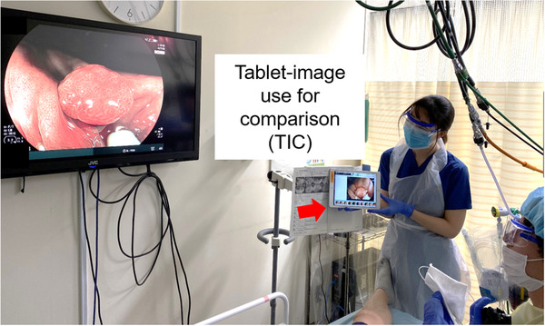
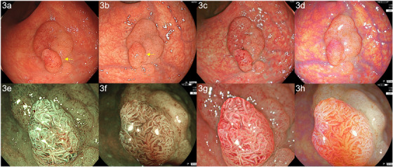
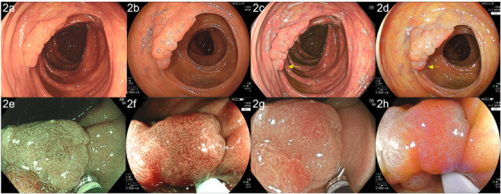
An endoscope system using 5-color light-emitting diodes (LEDs) (EVIS X1: CV-1500, Olympus Co., Tokyo, Japan) was released worldwide in July 2020. In addition to the improvement of narrow band imaging (NBI), this system enables texture and color enhancement imaging (TXI). TXI makes the lesion reddish and supports better visibility of colorectal lesions in comparison to white light imaging for improving lesion detection. On the other hand, another 4-color LED endoscope system (ELUXEO: BL-7000; Fujifilm, Tokyo, Japan) has been on the market in the West since 2017. This system enables blue light imaging (BLI) and linked color imaging (LCI). Generally, the accurate comparison between two images obtained by two different endoscope systems is difficult. To resolve this problem, we developed a method named the tablet-image comparison (TIC) method. TIC is a simple, easy, and paperless method to get images under similar conditions of two endoscope systems for an accurate comparison. We herein report two colorectal lesions in which accurate comparisons of images between TXI and LCI and between improved NBI and BLI obtained in the EVIS X1 and ELUXEO systems were performed using the TIC method. One was IIa 30 mm (high-grade dysplasia) and the other was IIa 25 mm (low-grade adenoma). A detailed comparison between TXI and LCI could be performed by TIC. In these two cases, with a distant view, TXI showed greater redness than LCI. LCI showed slightly higher brightness than TXI. In magnified TXI and LCI, the irregularities observed were similar to NBI and BLI, respectively.
一款采用5色发光二极管(LED)的内窥镜系统(EVIS X1:CV - 1500,日本东京奥林巴斯公司)于2020年7月在全球发布。除了窄带成像(NBI)有所改进外,该系统还具备纹理和色彩增强成像(TXI)功能。与白光成像相比,TXI能使病变呈现红色,有助于更好地观察结直肠病变,从而提高病变检测能力。另一方面,另一款4色LED内窥镜系统(ELUXEO:BL - 7000;日本东京富士胶片公司)自2017年起在西方市场上市。该系统具备蓝光成像(BLI)和联动彩色成像(LCI)功能。一般来说,准确比较由两种不同内窥镜系统获取的两幅图像是困难的。为解决这一问题,我们开发了一种名为平板图像比较(TIC)的方法。TIC是一种简单、便捷且无纸化的方法,可在相似条件下获取两种内窥镜系统的图像以进行准确比较。在此,我们报告两例结直肠病变,使用TIC方法对EVIS X1和ELUXEO系统中获得的TXI与LCI以及改进后的NBI与BLI之间的图像进行了准确比较。一例为30mm的IIa型(高级别异型增生),另一例为25mm的IIa型(低级别腺瘤)。通过TIC可以对TXI和LCI进行详细比较。在这两例中,从远处观察,TXI显示出比LCI更红的颜色。LCI的亮度略高于TXI。在放大的TXI和LCI中,观察到的不规则情况分别与NBI和BLI相似。