Department of Biochemistry and Molecular Biology, University of Kansas Medical Center, Kansas City, KS 66160, USA.
Zilkha Neurogenetic Institute, University of Southern California, Los Angeles, CA 90033, USA.
Biochim Biophys Acta Proteins Proteom. 2019 Jul-Aug;1867(7-8):691-700. doi: 10.1016/j.bbapap.2019.04.006. Epub 2019 Apr 18.
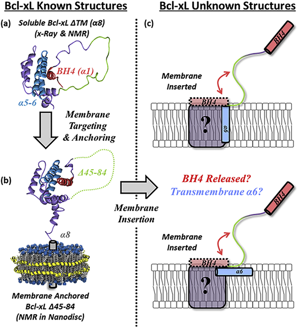
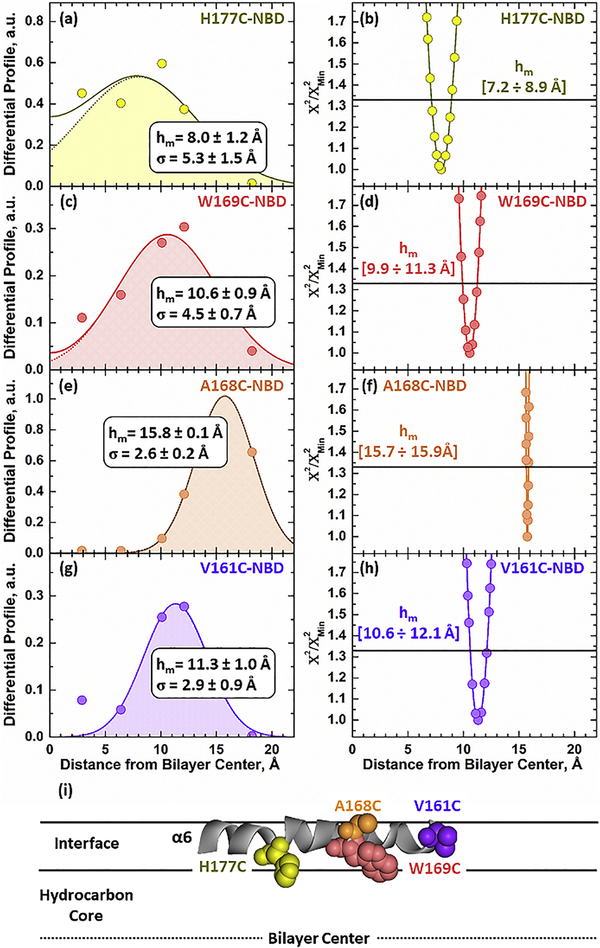
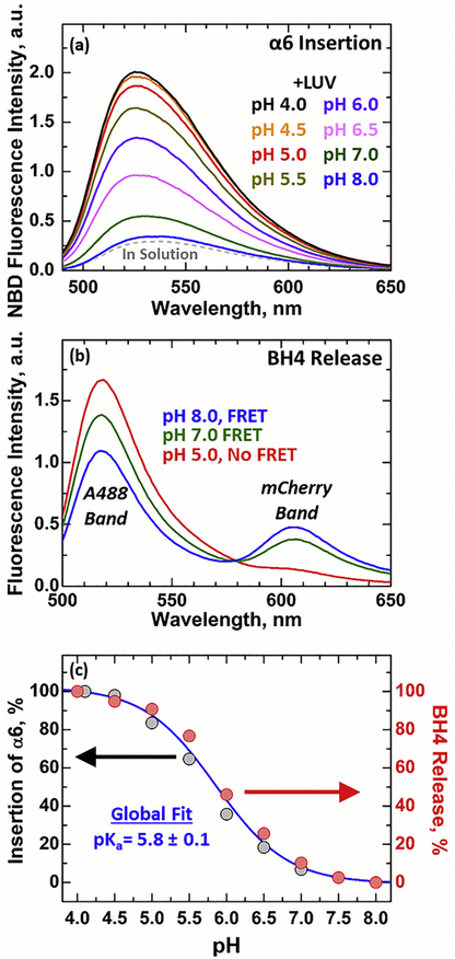
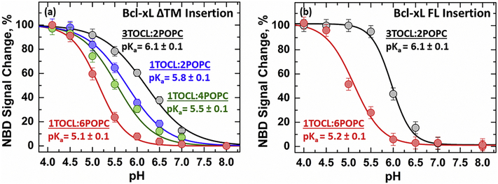
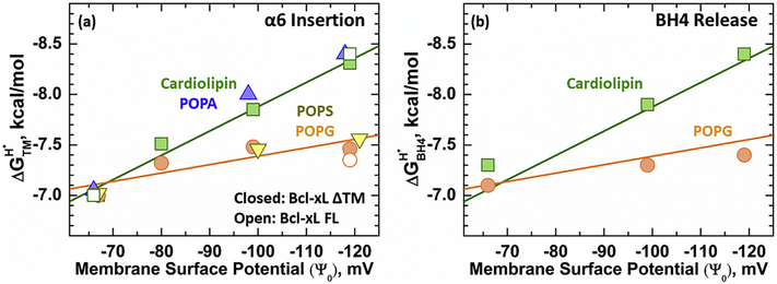
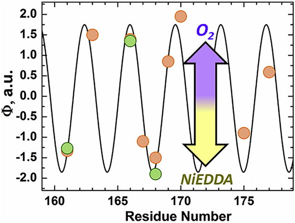
Bcl-xL is a member of the Bcl-2 family of apoptotic regulators, responsible for inhibiting the permeabilization of the mitochondrial outer membrane, and a promising anti-cancer target. Bcl-xL exists in the following conformations, each believed to play a role in the inhibition of apoptosis: (a) a soluble folded conformation, (b) a membrane-anchored (by its C-terminal α8 helix) form, which retains the same fold as in solution and (c) refolded membrane-inserted conformations, for which no structural data are available. Previous studies established that in the cell Bcl-xL exists in a dynamic equilibrium between soluble and membranous states, however, no direct evidence exists in support of either anchored or inserted conformation of the membranous state in vivo. In this in vitro study, we employed a combination of fluorescence and EPR spectroscopy to characterize structural features of the bilayer-inserted conformation of Bcl-xL and the lipid modulation of its membrane insertion transition. Our results indicate that the core hydrophobic helix α6 inserts into the bilayer without adopting a transmembrane orientation. This insertion disrupts the packing of Bcl-xL and releases the regulatory N-terminal BH4 domain (α1) from the rest of the protein structure. Our data demonstrate that both insertion and refolding of Bcl-xL are modulated by lipid composition, which brings the apparent pK of insertion to the threshold of physiological pH. We hypothesize that conformational rearrangements associated with the bilayer insertion of Bcl-xL result in its switching to a so-called non-canonical mode of apoptotic inhibition. Presented results suggest that the alteration in lipid composition before and during apoptosis can serve as an additional factor regulating the permeabilization of the mitochondrial outer membrane.
Bcl-xL 是凋亡调节因子 Bcl-2 家族的成员,负责抑制线粒体外膜的通透性,是一种很有前途的抗癌靶点。Bcl-xL 存在以下构象,每种构象都被认为在抑制细胞凋亡中发挥作用:(a) 可溶性折叠构象,(b) 膜锚定(通过其 C 末端α8 螺旋)形式,其保留与溶液中相同的折叠,(c) 重新折叠的膜插入构象,目前尚无结构数据。先前的研究表明,在细胞中,Bcl-xL 存在于可溶性和膜性状态之间的动态平衡,但没有直接证据支持膜性状态下的锚定或插入构象。在这项体外研究中,我们采用荧光和 EPR 光谱学相结合的方法来研究 Bcl-xL 的双层插入构象的结构特征及其膜插入转变的脂质调节。我们的结果表明,核心疏水性α6 螺旋插入双层,而不采用跨膜取向。这种插入破坏了 Bcl-xL 的包装,并将调节性 N 端 BH4 结构域(α1)从蛋白质结构的其余部分释放出来。我们的数据表明,Bcl-xL 的插入和重折叠均受脂质组成的调节,这将插入的表观 pK 值带到生理 pH 值的阈值。我们假设与 Bcl-xL 双层插入相关的构象重排导致其切换到所谓的非经典凋亡抑制模式。目前的结果表明,凋亡前和凋亡过程中脂质组成的改变可以作为调节线粒体外膜通透性的另一个因素。