Instituto de Microbiologia Paulo de Góes, Universidade Federal do Rio de Janeiro, Rio de Janeiro, Brazil.
Instituto de Ciências Biomédicas, Universidade Federal do Rio de Janeiro, Rio de Janeiro, Brazil.
PLoS One. 2019 Apr 23;14(4):e0215657. doi: 10.1371/journal.pone.0215657. eCollection 2019.
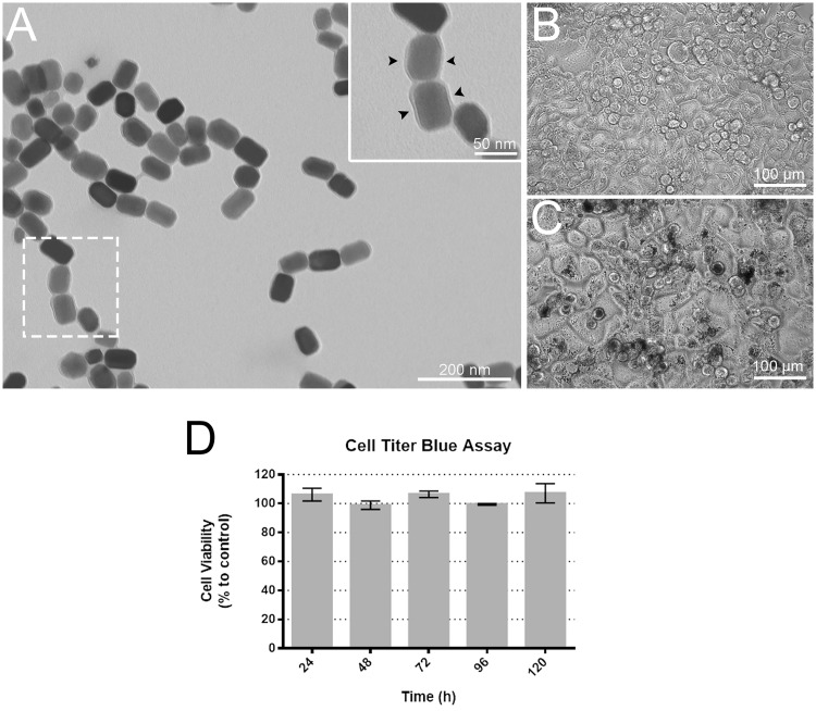
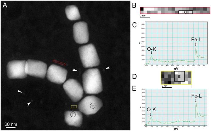
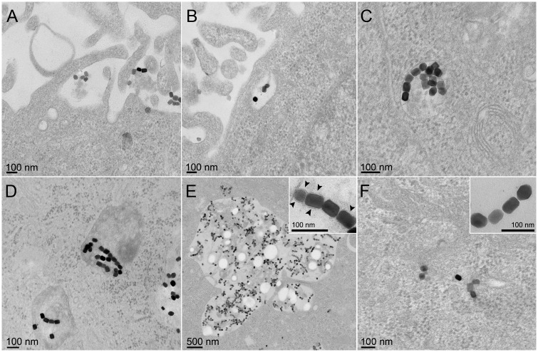
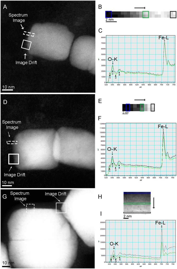
Magnetotactic bacteria biomineralize intracellular magnetic nanocrystals surrounded by a lipid bilayer called magnetosomes. Due to their unique characteristics, magnetite magnetosomes are promising tools in Biomedicine. However, the uptake, persistence, and accumulation of magnetosomes within mammalian cells have not been well studied. Here, the endocytic pathway of magnetite magnetosomes and their effects on human cervix epithelial (HeLa) cells were studied by electron microscopy and high spatial resolution nano-analysis techniques. Transmission electron microscopy of HeLa cells after incubation with purified magnetosomes showed the presence of magnetic nanoparticles inside or outside endosomes within the cell, which suggests different modes of internalization, and that these structures persisted beyond 120 h after internalization. High-resolution transmission electron microscopy and electron energy loss spectra of internalized magnetosome crystals showed no structural or chemical changes in these structures. Although crystal morphology was preserved, iron oxide crystalline particles of approximately 5 nm near internalized magnetosomes suggests that minor degradation of the original mineral structures might occur. Cytotoxicity and microscopy analysis showed that magnetosomes did not result in any apparent effect on HeLa cells viability or morphology. Based on our results, magnetosomes have significant biocompatibility with mammalian cells and thus have great potential in medical, biotechnological applications.
趋磁细菌生物矿化内部的磁性纳米晶体,被称为磁小体的脂质双层所包围。由于其独特的特性,磁铁矿磁小体是生物医学中很有前途的工具。然而,磁铁矿磁小体在哺乳动物细胞内的摄取、持久性和积累尚未得到很好的研究。在这里,通过电子显微镜和高空间分辨率纳米分析技术研究了磁铁矿磁小体的内吞途径及其对人宫颈上皮(HeLa)细胞的影响。用纯化的磁小体孵育后的 HeLa 细胞的透射电子显微镜显示,细胞内的内体中存在磁性纳米颗粒,这表明存在不同的内化方式,并且这些结构在内化后 120 小时仍然存在。内化的磁小体晶体的高分辨率透射电子显微镜和电子能量损失谱显示这些结构没有结构或化学变化。虽然晶体形态得以保留,但在内部化的磁小体附近存在约 5nm 的氧化铁晶状颗粒表明原始矿物结构可能发生了轻微降解。细胞毒性和显微镜分析表明,磁小体对 HeLa 细胞活力或形态没有明显影响。基于我们的结果,磁小体与哺乳动物细胞具有显著的生物相容性,因此在医学、生物技术应用中有很大的潜力。