Department of Physics, University of Cambridge, JJ Thomson Avenue, Cambridge, CB3 0HE, UK.
Cancer Research UK Cambridge Institute, University of Cambridge, Li Ka Shing Centre, Cambridge, CB2 0RE, UK.
Nat Commun. 2019 Apr 23;10(1):1902. doi: 10.1038/s41467-019-09484-4.
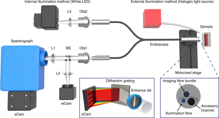
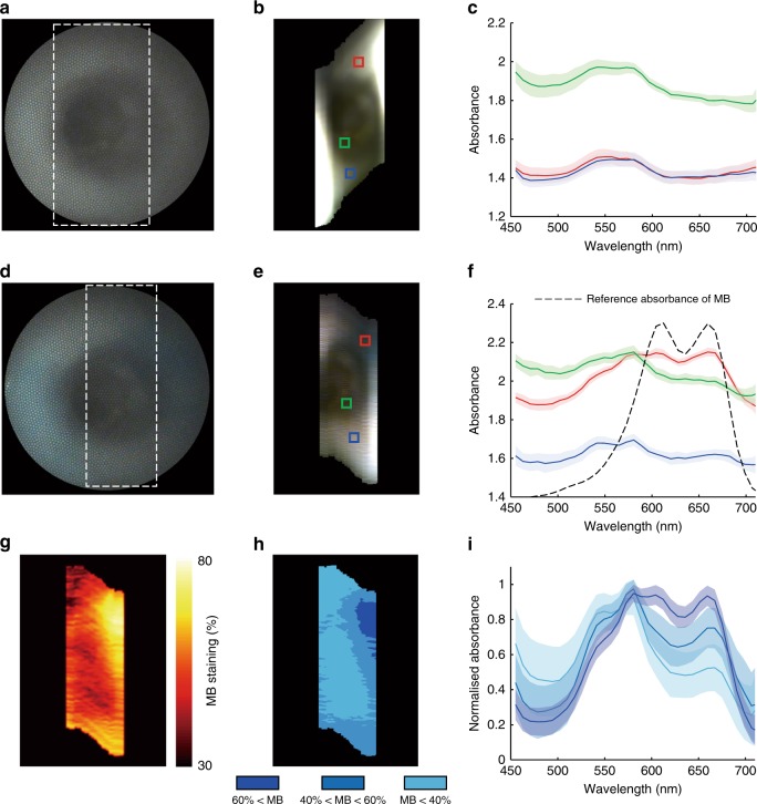
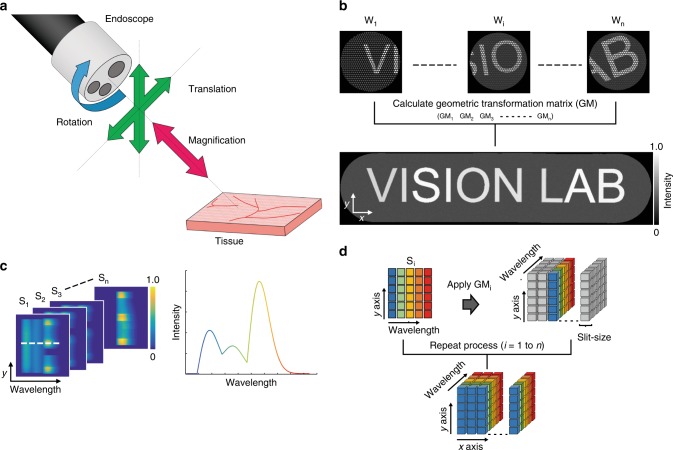
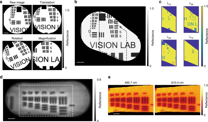
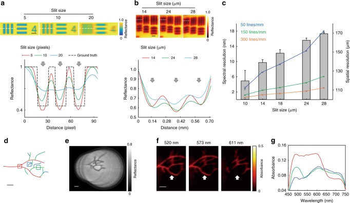
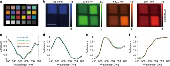
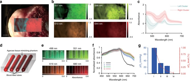
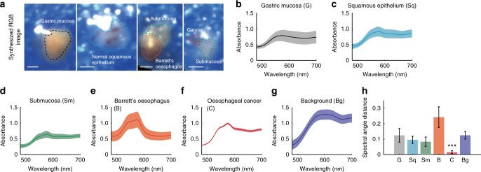
Hyperspectral imaging (HSI) enables visualisation of morphological and biochemical information, which could improve disease diagnostic accuracy. Unfortunately, the wide range of image distortions that arise during flexible endoscopy in the clinic have made integration of HSI challenging. To address this challenge, we demonstrate a hyperspectral endoscope (HySE) that simultaneously records intrinsically co-registered hyperspectral and standard-of-care white light images, which allows image distortions to be compensated computationally and an accurate hyperspectral data cube to be reconstructed as the endoscope moves in the lumen. Evaluation of HySE performance shows excellent spatial, spectral and temporal resolution and high colour fidelity. Application of HySE enables: quantification of blood oxygenation levels in tissue mimicking phantoms; differentiation of spectral profiles from normal and pathological ex vivo human tissues; and recording of hyperspectral data under freehand motion within an intact ex vivo pig oesophagus model. HySE therefore shows potential for enabling HSI in clinical endoscopy.
高光谱成象(HSI)使形态和生化信息可视化成为可能,这可能提高疾病诊断的准确性。不幸的是,在临床中进行柔性内窥镜检查时出现的广泛的图像变形使得 HSI 的集成具有挑战性。为了解决这个挑战,我们展示了一种高光谱内窥镜(HySE),它可以同时记录固有共配准的高光谱和标准护理白光图像,这允许通过计算来补偿图像变形,并在内窥镜在管腔中移动时重建准确的高光谱数据立方体。HySE 性能的评估表明具有出色的空间、光谱和时间分辨率以及高色彩保真度。HySE 的应用可以实现:在组织模拟体模中定量血氧水平;从正常和病理离体人组织中区分光谱曲线;以及在完整的离体猪食道模型中进行徒手运动下的高光谱数据记录。因此,HySE 有可能在临床内窥镜检查中实现 HSI。