• 文献检索
  • 文档翻译
  • 深度研究
  • 学术资讯
  • Suppr Zotero 插件Zotero 插件
  • 邀请有礼
  • 套餐&价格
  • 历史记录
应用&插件
Suppr Zotero 插件Zotero 插件浏览器插件Mac 客户端Windows 客户端微信小程序
定价
高级版会员购买积分包购买API积分包
服务
文献检索文档翻译深度研究API 文档MCP 服务
关于我们
关于 Suppr公司介绍联系我们用户协议隐私条款
关注我们

Suppr 超能文献

核心技术专利:CN118964589B侵权必究
粤ICP备2023148730 号-1Suppr @ 2026

文献检索

告别复杂PubMed语法,用中文像聊天一样搜索,搜遍4000万医学文献。AI智能推荐,让科研检索更轻松。

立即免费搜索

文件翻译

保留排版,准确专业,支持PDF/Word/PPT等文件格式,支持 12+语言互译。

免费翻译文档

深度研究

AI帮你快速写综述,25分钟生成高质量综述,智能提取关键信息,辅助科研写作。

立即免费体验

自动化荧光显微镜图像分割以用于人源性心肌球 3D 细胞检测。

Automated Segmentation of Fluorescence Microscopy Images for 3D Cell Detection in human-derived Cardiospheres.

机构信息

Department of Electronics and Telecommunications, Politecnico di Torino, Turin, 10129, Italy.

Department of Mechanical and Aerospace Engineering, Politecnico di Torino, Turin, 10129, Italy.

出版信息

Sci Rep. 2019 Apr 30;9(1):6644. doi: 10.1038/s41598-019-43137-2.

DOI:10.1038/s41598-019-43137-2
PMID:31040327
原文链接:https://pmc.ncbi.nlm.nih.gov/articles/PMC6491482/
Abstract

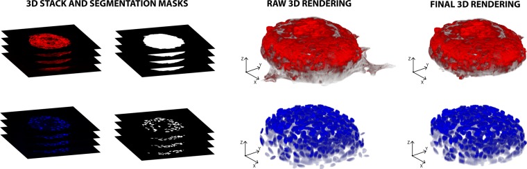
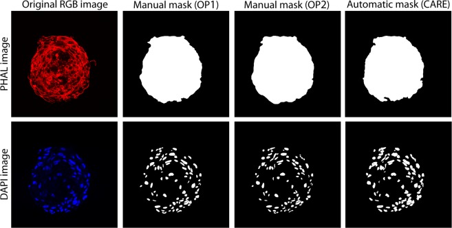
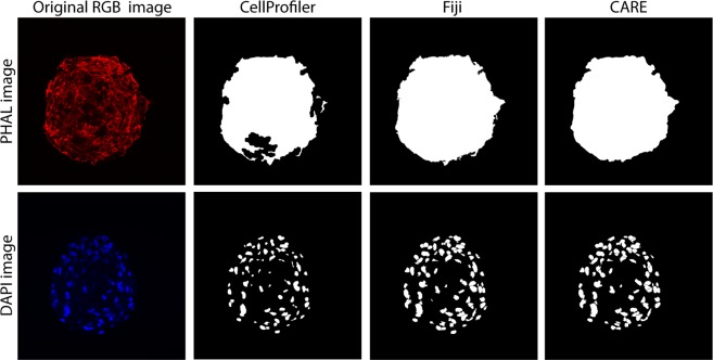
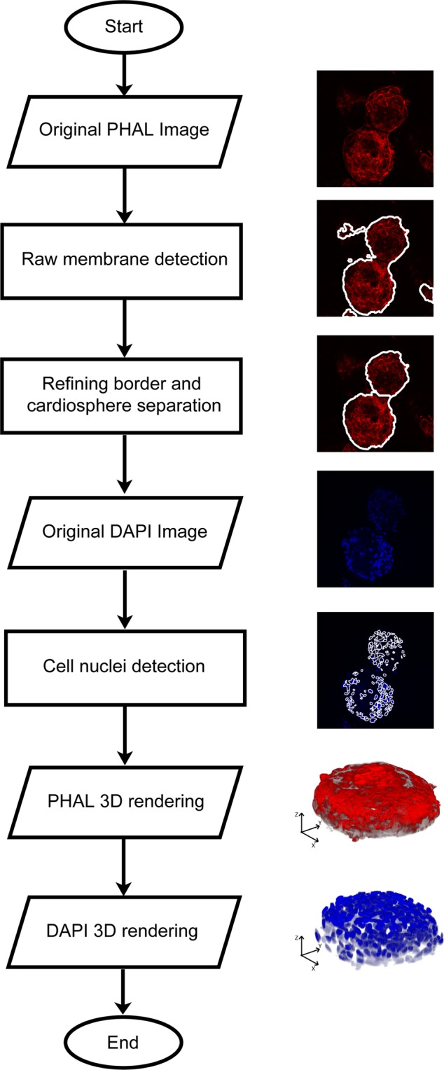
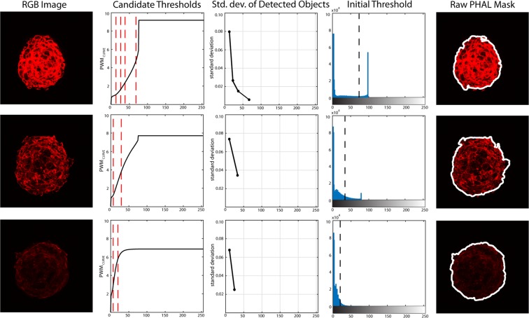
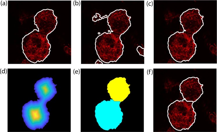
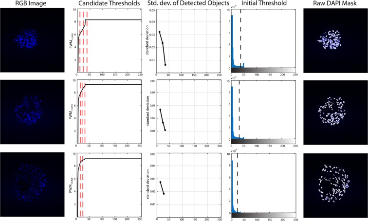
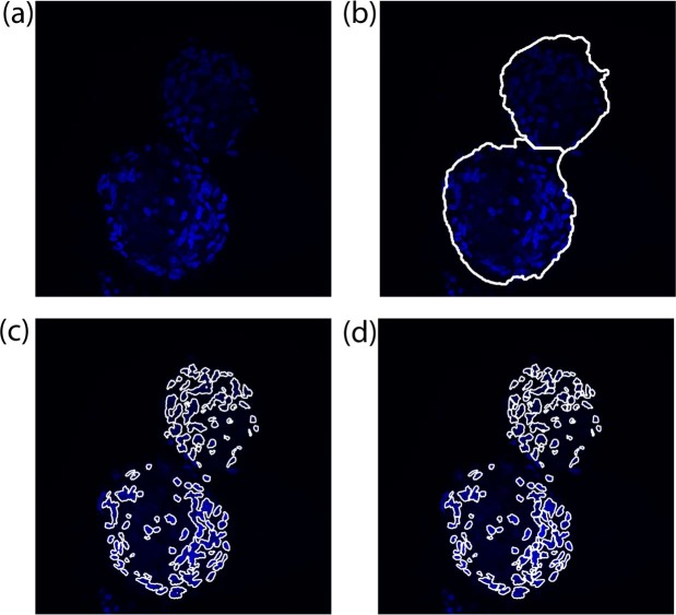
The 'cardiosphere' is a 3D cluster of cardiac progenitor cells recapitulating a stem cell niche-like microenvironment with a potential for disease and regeneration modelling of the failing human myocardium. In this multicellular 3D context, it is extremely important to decrypt the spatial distribution of cell markers for dissecting the evolution of cellular phenotypes by direct quantification of fluorescent signals in confocal microscopy. In this study, we present a fully automated method, named CARE ('CARdiosphere Evaluation'), for the segmentation of membranes and cell nuclei in human-derived cardiospheres. The proposed method is tested on twenty 3D-stacks of cardiospheres, for a total of 1160 images. Automatic results are compared with manual annotations and two open-source software designed for fluorescence microscopy. CARE performance was excellent in cardiospheres membrane segmentation and, in cell nuclei detection, the algorithm achieved the same performance as two expert operators. To the best of our knowledge, CARE is the first fully automated algorithm for segmentation inside in vitro 3D cell spheroids, including cardiospheres. The proposed approach will provide, in the future, automated quantitative analysis of markers distribution within the cardiac niche-like environment, enabling predictive associations between cell mechanical stresses and dynamic phenotypic changes.

摘要

“心脏球体”是一种 3D 簇状的心脏祖细胞,可再现具有潜在疾病和衰竭人类心肌再生建模能力的干细胞样微环境。在这个多细胞 3D 环境中,通过共聚焦显微镜中荧光信号的直接定量,解析细胞标记物的空间分布对于剖析细胞表型的演变非常重要。在这项研究中,我们提出了一种名为 CARE(“心脏球体评估”)的全自动方法,用于分割人类来源的心脏球体中的细胞膜和细胞核。该方法在 20 个心脏球体的 3D 堆栈上进行了测试,总共 1160 张图像。自动结果与手动注释和两个专为荧光显微镜设计的开源软件进行了比较。CARE 在心脏球体的细胞膜分割中表现出色,在细胞核检测中,该算法与两名专家操作人员的表现相同。据我们所知,CARE 是第一个用于分割体外 3D 细胞球体(包括心脏球体)内部的全自动算法。该方法将为心脏样环境中标记物分布的自动定量分析提供支持,从而能够预测细胞机械应力与动态表型变化之间的关联。

https://cdn.ncbi.nlm.nih.gov/pmc/blobs/173b/6491482/4e99d6a1d899/41598_2019_43137_Fig9_HTML.jpg
https://cdn.ncbi.nlm.nih.gov/pmc/blobs/173b/6491482/5f95dfb612d4/41598_2019_43137_Fig1_HTML.jpg
https://cdn.ncbi.nlm.nih.gov/pmc/blobs/173b/6491482/1b92872efc6b/41598_2019_43137_Fig2_HTML.jpg
https://cdn.ncbi.nlm.nih.gov/pmc/blobs/173b/6491482/26ec61259f9e/41598_2019_43137_Fig3_HTML.jpg
https://cdn.ncbi.nlm.nih.gov/pmc/blobs/173b/6491482/ee3d92a96505/41598_2019_43137_Fig4_HTML.jpg
https://cdn.ncbi.nlm.nih.gov/pmc/blobs/173b/6491482/47dfabfc6e51/41598_2019_43137_Fig5_HTML.jpg
https://cdn.ncbi.nlm.nih.gov/pmc/blobs/173b/6491482/854e73287338/41598_2019_43137_Fig6_HTML.jpg
https://cdn.ncbi.nlm.nih.gov/pmc/blobs/173b/6491482/c4f3d94fabc8/41598_2019_43137_Fig7_HTML.jpg
https://cdn.ncbi.nlm.nih.gov/pmc/blobs/173b/6491482/6a733a0be928/41598_2019_43137_Fig8_HTML.jpg
https://cdn.ncbi.nlm.nih.gov/pmc/blobs/173b/6491482/4e99d6a1d899/41598_2019_43137_Fig9_HTML.jpg

相似文献

1
Automated Segmentation of Fluorescence Microscopy Images for 3D Cell Detection in human-derived Cardiospheres.自动化荧光显微镜图像分割以用于人源性心肌球 3D 细胞检测。
Sci Rep. 2019 Apr 30;9(1):6644. doi: 10.1038/s41598-019-43137-2.
2
Automatic three-dimensional segmentation of mouse embryonic stem cell nuclei by utilising multiple channels of confocal fluorescence images.利用共聚焦荧光图像的多个通道自动对小鼠胚胎干细胞核进行三维分割。
J Microsc. 2021 Jan;281(1):57-75. doi: 10.1111/jmi.12949. Epub 2020 Aug 8.
3
FocAn: automated 3D analysis of DNA repair foci in image stacks acquired by confocal fluorescence microscopy.FocAn:通过共聚焦荧光显微镜获取的图像堆栈中 DNA 修复焦点的自动 3D 分析。
BMC Bioinformatics. 2020 Jan 28;21(1):27. doi: 10.1186/s12859-020-3370-8.
4
An analytical tool that quantifies cellular morphology changes from three-dimensional fluorescence images.一种可对三维荧光图像中的细胞形态变化进行量化的分析工具。
J Vis Exp. 2012 Aug 31(66):e4233. doi: 10.3791/4233.
5
Automated segmentation of brain cells for clonal analyses in fluorescence microscopy images.荧光显微镜图像中用于克隆分析的脑细胞自动分割。
J Neurosci Methods. 2019 Sep 1;325:108348. doi: 10.1016/j.jneumeth.2019.108348. Epub 2019 Jul 5.
6
Robust and automated three-dimensional segmentation of densely packed cell nuclei in different biological specimens with Lines-of-Sight decomposition.利用视线分解对不同生物样本中紧密堆积的细胞核进行稳健且自动化的三维分割。
BMC Bioinformatics. 2015 Jun 8;16:187. doi: 10.1186/s12859-015-0617-x.
7
PIWI-interacting RNA (piRNA) signatures in human cardiac progenitor cells.人类心脏祖细胞中的PIWI相互作用RNA(piRNA)特征
Int J Biochem Cell Biol. 2016 Jul;76:1-11. doi: 10.1016/j.biocel.2016.04.012. Epub 2016 Apr 27.
8
Ellipsoid Segmentation Model for Analyzing Light-Attenuated 3D Confocal Image Stacks of Fluorescent Multi-Cellular Spheroids.用于分析荧光多细胞球体光衰减3D共聚焦图像堆栈的椭球体分割模型
PLoS One. 2016 Jun 15;11(6):e0156942. doi: 10.1371/journal.pone.0156942. eCollection 2016.
9
Quantitative Analysis of Whole-Mount Fluorescence-Stained Tumor Spheroids in Phenotypic Drug Screens.全荧光染色肿瘤球体在表型药物筛选中的定量分析。
Methods Mol Biol. 2024;2764:311-334. doi: 10.1007/978-1-0716-3674-9_20.
10
AnaSP: a software suite for automatic image analysis of multicellular spheroids.AnaSP:用于多细胞球体自动图像分析的软件套件。
Comput Methods Programs Biomed. 2015 Apr;119(1):43-52. doi: 10.1016/j.cmpb.2015.02.006. Epub 2015 Feb 24.

引用本文的文献

1
Harnessing 3D cell models and high-resolution imaging to unveil the mechanisms of nanoparticle-mediated drug delivery.利用3D细胞模型和高分辨率成像揭示纳米颗粒介导的药物递送机制。
Front Bioeng Biotechnol. 2025 Jul 7;13:1606573. doi: 10.3389/fbioe.2025.1606573. eCollection 2025.
2
Blockade of YAP Mechanoactivation Prevents Neointima Formation and Adverse Remodeling in Arterialized Vein Grafts.YAP 机械激活的阻断可预防动脉化静脉移植物中的新生内膜形成和不良重塑。
J Am Heart Assoc. 2025 Apr;14(7):e037531. doi: 10.1161/JAHA.124.037531. Epub 2025 Mar 21.
3
Comparing Deep Learning Performance for Chronic Lymphocytic Leukaemia Cell Segmentation in Brightfield Microscopy Images.

本文引用的文献

1
Multi-tissue and multi-scale approach for nuclei segmentation in H&E stained images.多组织和多尺度方法用于 H&E 染色图像中的细胞核分割。
Biomed Eng Online. 2018 Jun 20;17(1):89. doi: 10.1186/s12938-018-0518-0.
2
Impact of physical confinement on nuclei geometry and cell division dynamics in 3D spheroids.物理限制对 3D 球体中核几何形状和细胞分裂动力学的影响。
Sci Rep. 2018 Jun 8;8(1):8785. doi: 10.1038/s41598-018-27060-6.
3
Modeling signaling-dependent pluripotency with Boolean logic to predict cell fate transitions.用布尔逻辑对信号依赖性多能性进行建模,以预测细胞命运转变。
比较深度学习在明场显微镜图像中对慢性淋巴细胞白血病细胞分割的性能。
Bioinform Biol Insights. 2024 Sep 5;18:11779322241272387. doi: 10.1177/11779322241272387. eCollection 2024.
4
Evaluation of Cellpose segmentation with sequential thresholding for instance segmentation of cytoplasms within autofluorescence images.评估 Cellpose 分割与顺序阈值法在自动荧光图像中细胞质实例分割的应用。
Comput Biol Med. 2024 Sep;179:108846. doi: 10.1016/j.compbiomed.2024.108846. Epub 2024 Jul 7.
5
Dopamine-mediated striatal activity and function is enhanced in GlyRα2 knockout animals.在甘氨酸受体α2基因敲除动物中,多巴胺介导的纹状体活动和功能增强。
iScience. 2023 Jul 17;26(8):107400. doi: 10.1016/j.isci.2023.107400. eCollection 2023 Aug 18.
6
Prevalence and practices of immunofluorescent cell image processing: a systematic review.免疫荧光细胞图像处理的患病率与实践:一项系统综述
Front Cell Neurosci. 2023 Jul 20;17:1188858. doi: 10.3389/fncel.2023.1188858. eCollection 2023.
7
POSEA: A novel algorithm to evaluate the performance of multi-object instance image segmentation.POSEA:一种用于评估多目标实例图像分割性能的新算法。
PLoS One. 2023 Mar 29;18(3):e0283692. doi: 10.1371/journal.pone.0283692. eCollection 2023.
8
Volumetric imaging of human mesenchymal stem cells (hMSCs) for non-destructive quantification of 3D cell culture growth.用于无损定量 3D 细胞培养生长的人骨髓间充质干细胞(hMSCs)容积成像。
PLoS One. 2023 Mar 28;18(3):e0282298. doi: 10.1371/journal.pone.0282298. eCollection 2023.
9
Segmentation of Tissues and Proliferating Cells in Light-Sheet Microscopy Images of Mouse Embryos Using Convolutional Neural Networks.使用卷积神经网络对小鼠胚胎光片显微镜图像中的组织和增殖细胞进行分割
IEEE Access. 2022;10:105084-105100. doi: 10.1109/access.2022.3210542. Epub 2022 Sep 28.
10
A comprehensive review of computational and image analysis techniques for quantitative evaluation of striated muscle tissue architecture.对用于横纹肌组织结构定量评估的计算和图像分析技术的全面综述。
Biophys Rev (Melville). 2022 Dec;3(4):041302. doi: 10.1063/5.0057434. Epub 2022 Nov 4.
Mol Syst Biol. 2018 Jan 29;14(1):e7952. doi: 10.15252/msb.20177952.
4
A stepwise model of reaction-diffusion and positional information governs self-organized human peri-gastrulation-like patterning.反应扩散和位置信息的逐步模型控制着自组织的人类原肠胚形成样模式。
Development. 2017 Dec 1;144(23):4298-4312. doi: 10.1242/dev.149658. Epub 2017 Sep 4.
5
Building bridges between cellular and molecular structural biology.搭建细胞与分子结构生物学之间的桥梁。
Elife. 2017 Jul 6;6:e25835. doi: 10.7554/eLife.25835.
6
Engineering cell fitness: lessons for regenerative medicine.工程细胞适应性:再生医学的启示。
Curr Opin Biotechnol. 2017 Oct;47:7-15. doi: 10.1016/j.copbio.2017.05.005. Epub 2017 May 25.
7
Dissecting the stem cell niche with organoid models: an engineering-based approach.利用类器官模型剖析干细胞生态位:一种基于工程学的方法。
Development. 2017 Mar 15;144(6):998-1007. doi: 10.1242/dev.140905.
8
Stem Cell Spheroids and Ex Vivo Niche Modeling: Rationalization and Scaling-Up.干细胞球体与体外生态位建模:合理化与放大
J Cardiovasc Transl Res. 2017 Apr;10(2):150-166. doi: 10.1007/s12265-017-9741-5. Epub 2017 Mar 13.
9
Put to the test.接受检验。
Elife. 2017 Jan 30;6:e24276. doi: 10.7554/eLife.24276.
10
Cardiac Mechanoperception: A Life-Long Story from Early Beats to Aging and Failure.心脏机械感知:从早期心跳到衰老与衰竭的一生故事。
Stem Cells Dev. 2017 Jan 15;26(2):77-90. doi: 10.1089/scd.2016.0206. Epub 2016 Nov 14.