• 文献检索
  • 文档翻译
  • 深度研究
  • 学术资讯
  • Suppr Zotero 插件Zotero 插件
  • 邀请有礼
  • 套餐&价格
  • 历史记录
应用&插件
Suppr Zotero 插件Zotero 插件浏览器插件Mac 客户端Windows 客户端微信小程序
定价
高级版会员购买积分包购买API积分包
服务
文献检索文档翻译深度研究API 文档MCP 服务
关于我们
关于 Suppr公司介绍联系我们用户协议隐私条款
关注我们

Suppr 超能文献

核心技术专利:CN118964589B侵权必究
粤ICP备2023148730 号-1Suppr @ 2026

文献检索

告别复杂PubMed语法,用中文像聊天一样搜索,搜遍4000万医学文献。AI智能推荐,让科研检索更轻松。

立即免费搜索

文件翻译

保留排版,准确专业,支持PDF/Word/PPT等文件格式,支持 12+语言互译。

免费翻译文档

深度研究

AI帮你快速写综述,25分钟生成高质量综述,智能提取关键信息,辅助科研写作。

立即免费体验

视网膜微血管的年龄相关性变化。

Age-related changes of the retinal microvasculature.

机构信息

Laboratory of Genetics & Genomics, National Institute on Aging/National Institutes of Health, Baltimore, Maryland, United States of America.

Division of Epidemiology and Clinical Applications, National Eye Institute/National Institutes of Health, Baltimore, Maryland, United States of America.

出版信息

PLoS One. 2019 May 2;14(5):e0215916. doi: 10.1371/journal.pone.0215916. eCollection 2019.

DOI:10.1371/journal.pone.0215916
PMID:31048908
原文链接:https://pmc.ncbi.nlm.nih.gov/articles/PMC6497255/
Abstract

PURPOSE

Blood vessels of the retina provide an easily-accessible, representative window into the condition of microvasculature. We investigated how retinal vessel structure captured in fundus photographs changes with age, and how this may reflect features related to patient health, including blood pressure.

RESULTS

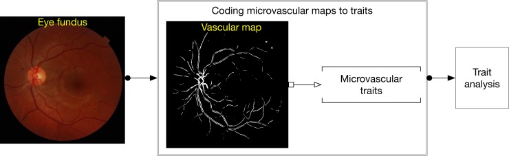
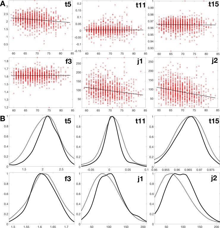
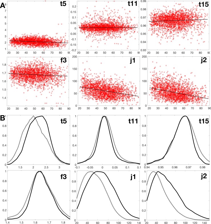
We used two approaches. In the first approach, we segmented the retinal vasculature from fundus photographs and then we correlated 25 parameterized aspects ("traits")-comprising 15 measures of tortuosity, 7 fractal ranges of self-similarity, and 3 measures of junction numbers-with participant age and blood pressure. In the second approach, we examined entire fundus photographs with a set of algorithmic CHARM features. We studied 2,280 Sardinians, ages 20-28, and an U.S. based population from the AREDS study in 1,178 participants, ages 59-84. Three traits (relating to tortuosity, vessel bifurcation number, and vessel endpoint number) showed significant changes with age in both cohorts, and one additional trait (relating to fractal number) showed a correlation in the Sardinian cohort only. When using second approach, we found significant correlations of particular CHARM features with age and blood pressure, which were stronger than those detected when using parameterized traits, reflecting a greater signal from the entire photographs than was captured in the segmented microvasculature.

CONCLUSIONS

These findings demonstrate that automated quantitative image analysis of fundus images can reveal general measures of patient health status.

摘要

目的

视网膜血管为了解微血管状况提供了一个易于接近的代表性窗口。我们研究了眼底照片中视网膜血管结构随年龄的变化,以及这种变化如何反映与患者健康相关的特征,包括血压。

结果

我们使用了两种方法。在第一种方法中,我们从眼底照片中分割出视网膜血管,然后将 25 个参数化方面(包括 15 个迂曲度测量、7 个自相似分形范围和 3 个连接点数测量)与参与者的年龄和血压进行相关分析。在第二种方法中,我们使用一组算法 CHARM 特征来检查整个眼底照片。我们研究了 2280 名年龄在 20-28 岁的撒丁岛人,以及 1178 名年龄在 59-84 岁的美国 AREDS 研究中的参与者。在两个队列中,有三个特征(与迂曲度、血管分叉数和血管终点数有关)随年龄发生显著变化,另外一个特征(与分形数有关)仅在撒丁岛队列中存在相关性。当使用第二种方法时,我们发现特定 CHARM 特征与年龄和血压之间存在显著相关性,其相关性强于使用参数化特征时的相关性,这反映了整个照片比分割后的微血管提供了更多的信号。

结论

这些发现表明,对眼底图像的自动定量图像分析可以揭示患者健康状况的一般指标。

https://cdn.ncbi.nlm.nih.gov/pmc/blobs/ebb3/6497255/c250b4673a21/pone.0215916.g003.jpg
https://cdn.ncbi.nlm.nih.gov/pmc/blobs/ebb3/6497255/d1b6281a8654/pone.0215916.g001.jpg
https://cdn.ncbi.nlm.nih.gov/pmc/blobs/ebb3/6497255/0d06a0ce146f/pone.0215916.g002.jpg
https://cdn.ncbi.nlm.nih.gov/pmc/blobs/ebb3/6497255/c250b4673a21/pone.0215916.g003.jpg
https://cdn.ncbi.nlm.nih.gov/pmc/blobs/ebb3/6497255/d1b6281a8654/pone.0215916.g001.jpg
https://cdn.ncbi.nlm.nih.gov/pmc/blobs/ebb3/6497255/0d06a0ce146f/pone.0215916.g002.jpg
https://cdn.ncbi.nlm.nih.gov/pmc/blobs/ebb3/6497255/c250b4673a21/pone.0215916.g003.jpg

相似文献

1
Age-related changes of the retinal microvasculature.视网膜微血管的年龄相关性变化。
PLoS One. 2019 May 2;14(5):e0215916. doi: 10.1371/journal.pone.0215916. eCollection 2019.
2
Retinal vascular fractal dimension is associated with cognitive dysfunction.视网膜血管分形维数与认知功能障碍有关。
J Stroke Cerebrovasc Dis. 2014 Jan;23(1):43-50. doi: 10.1016/j.jstrokecerebrovasdis.2012.09.002. Epub 2012 Oct 23.
3
Morphological Analysis of Retinal Microvasculature to Improve Understanding of Retinal Hemorrhage Mechanics in Infants.分析视网膜微血管形态,加深对婴儿期视网膜出血力学的理解。
Invest Ophthalmol Vis Sci. 2020 Mar 9;61(3):16. doi: 10.1167/iovs.61.3.16.
4
Age-related decrease of the retinal vasculature area identified with a novel computer-aided analysis system.新型计算机辅助分析系统识别的与年龄相关的视网膜血管面积减少。
Tohoku J Exp Med. 2012 Nov;228(3):229-37. doi: 10.1620/tjem.228.229.
5
Revised formulas for summarizing retinal vessel diameters.用于总结视网膜血管直径的修订公式。
Curr Eye Res. 2003 Sep;27(3):143-9. doi: 10.1076/ceyr.27.3.143.16049.
6
The retinal vasculature as a fractal: methodology, reliability, and relationship to blood pressure.视网膜血管系统作为一种分形结构:方法、可靠性及其与血压的关系。
Ophthalmology. 2008 Nov;115(11):1951-6. doi: 10.1016/j.ophtha.2008.05.029. Epub 2008 Aug 9.
7
Ocular Fundus Photography as a Tool to Study Stroke and Dementia.眼底摄影作为研究中风和痴呆的工具。
Semin Neurol. 2015 Oct;35(5):481-90. doi: 10.1055/s-0035-1563570. Epub 2015 Oct 6.
8
[A study on automatic and quantitative diagnosis of fundus photographs. I. Detection of contour line of retinal blood vessel images on color fundus photographs (author's transl)].[眼底照片自动定量诊断的研究。I. 彩色眼底照片上视网膜血管图像轮廓线的检测(作者译)]
Nippon Ganka Gakkai Zasshi. 1973 Aug;77(8):907-18.
9
Retinal blood vessel width measured on color fundus photographs by image analysis.通过图像分析在彩色眼底照片上测量视网膜血管宽度。
Acta Ophthalmol Scand Suppl. 1995(215):33-40. doi: 10.1111/j.1600-0420.1995.tb00590.x.
10
A spectrum of retinal vasculature measures and coronary artery disease.视网膜血管测量与冠状动脉疾病的关系。
Atherosclerosis. 2018 Jan;268:215-224. doi: 10.1016/j.atherosclerosis.2017.10.008. Epub 2017 Oct 10.

引用本文的文献

1
Characterizing Retinal Mitochondrial Function and Its Relations to Intraretinal Thicknesses in Healthy Adults.健康成年人视网膜线粒体功能及其与视网膜内厚度的关系特征
Curr Eye Res. 2025 Jul 9:1-8. doi: 10.1080/02713683.2025.2527755.
2
Convolutional Neural Network-Based Prediction of Axial Length Using Color Fundus Photography.基于卷积神经网络的彩色眼底照相轴向长度预测。
Transl Vis Sci Technol. 2024 May 1;13(5):23. doi: 10.1167/tvst.13.5.23.
3
Understanding human aging and the fundamental cell signaling link in age-related diseases: the middle-aging hypovascularity hypoxia hypothesis.

本文引用的文献

1
Prediction of cardiovascular risk factors from retinal fundus photographs via deep learning.基于深度学习的眼底图像心血管风险因素预测。
Nat Biomed Eng. 2018 Mar;2(3):158-164. doi: 10.1038/s41551-018-0195-0. Epub 2018 Feb 19.
2
Association between the retinal vascular network with Singapore "I" Vessel Assessment (SIVA) software, cardiovascular history and risk factors in the elderly: The Montrachet study, population-based study.老年人视网膜血管网络与新加坡“I”血管评估 (SIVA) 软件、心血管病史和危险因素的关系:基于人群的蒙丽榭研究。
PLoS One. 2018 Apr 3;13(4):e0194694. doi: 10.1371/journal.pone.0194694. eCollection 2018.
3
理解人类衰老以及与年龄相关疾病中的基本细胞信号传导联系:中年血管减少性缺氧假说。
Front Aging. 2023 Jun 13;4:1196648. doi: 10.3389/fragi.2023.1196648. eCollection 2023.
4
Assessment of factors affecting flicker ERGs recorded with RETeval from data obtained from health checkup screening.评估影响从健康检查筛查中获得的数据记录的闪烁视网膜电图(flicker ERGs)的因素。
PLoS One. 2023 Apr 24;18(4):e0284686. doi: 10.1371/journal.pone.0284686. eCollection 2023.
5
Repeatability of Peripapillary Optical Coherence Tomography Angiography Parameters in Older Adults.老年人视乳头周围光学相干断层扫描血管造影参数的可重复性
J Vitreoretin Dis. 2020 Oct 1;5(3):239-246. doi: 10.1177/2474126420953968. eCollection 2021 May-Jun.
6
The Role of Oxidative Stress in the Aging Eye.氧化应激在衰老眼睛中的作用。
Life (Basel). 2023 Mar 20;13(3):837. doi: 10.3390/life13030837.
7
An Analysis of Metabolic Changes in the Retina and Retinal Pigment Epithelium of Aging Mice.衰老小鼠视网膜及视网膜色素上皮细胞代谢变化分析
Invest Ophthalmol Vis Sci. 2021 Nov 1;62(14):20. doi: 10.1167/iovs.62.14.20.
8
Age-Related Retinal Changes in Wild-Type C57BL/6J Mice Between 2 and 32 Months.2 至 32 月龄 C57BL/6J 野生型小鼠的年龄相关性视网膜变化。
Invest Ophthalmol Vis Sci. 2021 Jun 1;62(7):9. doi: 10.1167/iovs.62.7.9.
9
Retinal microvascular parameters are not significantly associated with mild cognitive impairment in the Northern Ireland Cohort for the Longitudinal Study of Ageing.北爱尔兰老龄化纵向研究队列中,视网膜微血管参数与轻度认知障碍无显著相关性。
BMC Neurol. 2021 Mar 11;21(1):112. doi: 10.1186/s12883-021-02137-4.
10
Association of reduced retinal arteriolar tortuosity with depression in older participants from the Northern Ireland Cohort for the Longitudinal Study of Ageing.与北爱尔兰老龄化纵向研究队列中年龄较大的参与者的抑郁相关的视网膜小动脉扭曲度降低。
BMC Geriatr. 2021 Jan 15;21(1):62. doi: 10.1186/s12877-021-02009-z.
Automated Grading of Age-Related Macular Degeneration From Color Fundus Images Using Deep Convolutional Neural Networks.
使用深度卷积神经网络从彩色眼底图像对年龄相关性黄斑变性进行自动分级
JAMA Ophthalmol. 2017 Nov 1;135(11):1170-1176. doi: 10.1001/jamaophthalmol.2017.3782.
4
Differential Aging Signals in Abdominal CT Scans.腹部CT扫描中的差异衰老信号
Acad Radiol. 2017 Dec;24(12):1535-1543. doi: 10.1016/j.acra.2017.07.007. Epub 2017 Sep 15.
5
Improved Automated Detection of Diabetic Retinopathy on a Publicly Available Dataset Through Integration of Deep Learning.通过深度学习整合在公开可用数据集上改进糖尿病视网膜病变的自动检测
Invest Ophthalmol Vis Sci. 2016 Oct 1;57(13):5200-5206. doi: 10.1167/iovs.16-19964.
6
Retinal Information is Independently Associated with Cardiovascular Disease in Patients with Type 2 diabetes.视网膜信息与2型糖尿病患者的心血管疾病独立相关。
Sci Rep. 2016 Jan 12;6:19053. doi: 10.1038/srep19053.
7
Retinal microvascular network attenuation in Alzheimer's disease.阿尔茨海默病中的视网膜微血管网络衰减
Alzheimers Dement (Amst). 2015 May 16;1(2):229-235. doi: 10.1016/j.dadm.2015.04.001. eCollection 2015 Jun.
8
Automatic identification of reticular pseudodrusen using multimodal retinal image analysis.使用多模态视网膜图像分析自动识别网状假性玻璃膜疣
Invest Ophthalmol Vis Sci. 2015 Jan 8;56(1):633-9. doi: 10.1167/iovs.14-15019.
9
Methods and reproducibility of grading optimized digital color fundus photographs in the Age-Related Eye Disease Study 2 (AREDS2 Report Number 2).年龄相关性眼病研究 2(AREDS2 报告编号 2)中优化的数字眼底彩色照片分级方法和可重复性。
Invest Ophthalmol Vis Sci. 2013 Jul 8;54(7):4548-54. doi: 10.1167/iovs.13-11804.
10
Retinal vascular fractal dimension is associated with cognitive dysfunction.视网膜血管分形维数与认知功能障碍有关。
J Stroke Cerebrovasc Dis. 2014 Jan;23(1):43-50. doi: 10.1016/j.jstrokecerebrovasdis.2012.09.002. Epub 2012 Oct 23.