Institute for Chemical and Physical Processes, IPCF, CNR, ss Pisa, Pisa, Italy.
IRCCS Cardiovascolare Multimedica, Milan, Italy.
J Tissue Eng Regen Med. 2019 Jul;13(7):1253-1264. doi: 10.1002/term.2875. Epub 2019 May 31.
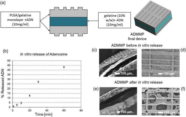
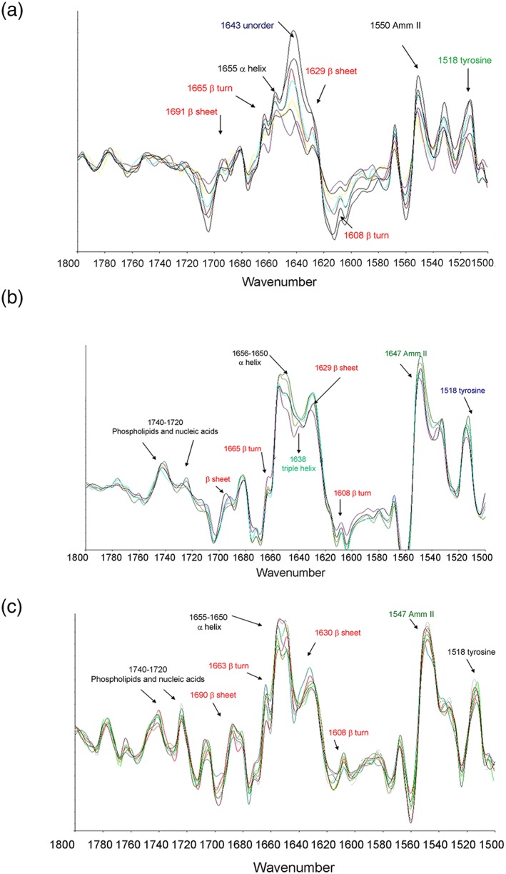
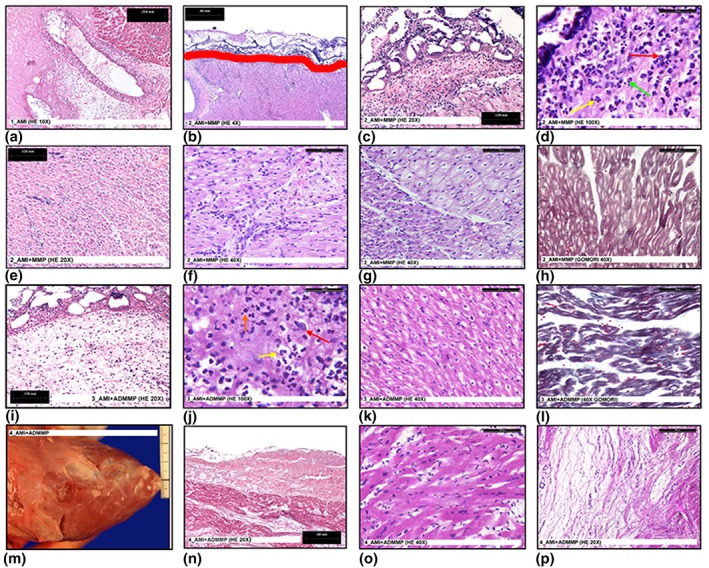
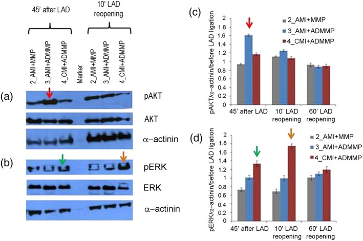
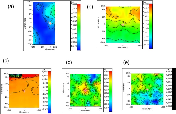
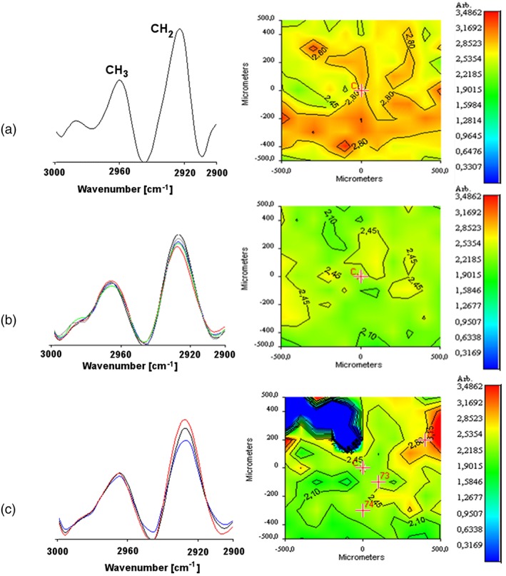
The protection from ischaemia-reperfusion-associated myocardial infarction worsening remains a big challenge. We produced a bioartificial 3D cardiac patch with cardioinductive properties on stem cells. Its multilayer structure was functionalised with clinically relevant doses of adenosine. We report here the first study on the potential of these cardiac patches in the controlled delivery of adenosine into the in vivo ischaemic-reperfused pig heart. A Fourier transform infrared chemical imaging approach allowed us to perform a characterisation, complementary to the histological and biochemical analyses on myocardial samples after in vivo patch implantation, increasing the number of investigations and results on the restricted number of pigs (n = 4) employed in this feasibility step. In vitro tests suggested that adenosine was completely released by a functionalised patch, a data that was confirmed in vivo after 24 hr from patch implantation. Moreover, the adenosine-loaded patch enabled a targeted delivery of the drug to the ischaemic-reperfused area of the heart, as highlighted by the activation of the pro-survival signalling reperfusion injury salvage kinases pathway. At 3 months, though limited to one animal, the used methods provided a picture of a tissue in dynamic conditions, associated to the biosynthesis of new collagen and to a non-fibrotic outcome of the healing process underway. The synergistic effect between the functionalised 3D cardiac patch and adenosine cardioprotection might represent a promising innovation in the treatment of reperfusion injury. As this is a feasibility study, the clinical implications of our findings will require further in vivo investigation on larger numbers of ischaemic-reperfused pig hearts.
防止缺血再灌注相关的心肌梗死恶化仍然是一个巨大的挑战。我们利用具有心脏诱导特性的干细胞制造了一种生物人工 3D 心脏贴片。其多层结构用临床相关剂量的腺苷进行了功能化。我们在这里报告了这些心脏贴片在体内缺血再灌注猪心脏中控制递腺苷的潜在用途的第一项研究。傅里叶变换红外化学成像方法使我们能够对心肌样本进行特征描述,这与体内贴片植入后的组织学和生化分析互补,增加了对这项可行性研究中有限数量(n=4)猪的研究和结果的数量。体外测试表明,功能化贴片可完全释放腺苷,该数据在贴片植入 24 小时后在体内得到证实。此外,载有腺苷的贴片能够将药物靶向递送到心脏的缺血再灌注区域,如再灌注损伤挽救激酶途径的存活信号的激活所强调的那样。尽管在 3 个月的时间里,只有一只动物被限制,但所使用的方法提供了一幅动态条件下的组织图像,与新胶原蛋白的生物合成以及正在进行的愈合过程的非纤维化结果相关。功能化 3D 心脏贴片和腺苷心脏保护的协同作用可能代表再灌注损伤治疗的一个有前途的创新。由于这是一项可行性研究,我们研究结果的临床意义需要在更多数量的缺血再灌注猪心脏上进行进一步的体内研究。