• 文献检索
  • 文档翻译
  • 深度研究
  • 学术资讯
  • Suppr Zotero 插件Zotero 插件
  • 邀请有礼
  • 套餐&价格
  • 历史记录
应用&插件
Suppr Zotero 插件Zotero 插件浏览器插件Mac 客户端Windows 客户端微信小程序
定价
高级版会员购买积分包购买API积分包
服务
文献检索文档翻译深度研究API 文档MCP 服务
关于我们
关于 Suppr公司介绍联系我们用户协议隐私条款
关注我们

Suppr 超能文献

核心技术专利:CN118964589B侵权必究
粤ICP备2023148730 号-1Suppr @ 2026

文献检索

告别复杂PubMed语法,用中文像聊天一样搜索,搜遍4000万医学文献。AI智能推荐,让科研检索更轻松。

立即免费搜索

文件翻译

保留排版,准确专业,支持PDF/Word/PPT等文件格式,支持 12+语言互译。

免费翻译文档

深度研究

AI帮你快速写综述,25分钟生成高质量综述,智能提取关键信息,辅助科研写作。

立即免费体验

一种用于从二维相位对比磁共振图像中进行稳健血流定量的新型血管分割算法。

A new vessel segmentation algorithm for robust blood flow quantification from two-dimensional phase-contrast magnetic resonance images.

作者信息

Bidhult Sebastian, Hedström Erik, Carlsson Marcus, Töger Johannes, Steding-Ehrenborg Katarina, Arheden Håkan, Aletras Anthony H, Heiberg Einar

机构信息

Department of Clinical Sciences Lund, Clinical Physiology, Skane University Hospital, Lund University, Lund, Sweden.

Department of Biomedical Engineering, Faculty of Engineering, Lund University, Lund, Sweden.

出版信息

Clin Physiol Funct Imaging. 2019 Sep;39(5):327-338. doi: 10.1111/cpf.12582. Epub 2019 Jun 6.

DOI:10.1111/cpf.12582
PMID:31102479
原文链接:https://pmc.ncbi.nlm.nih.gov/articles/PMC6852024/
Abstract

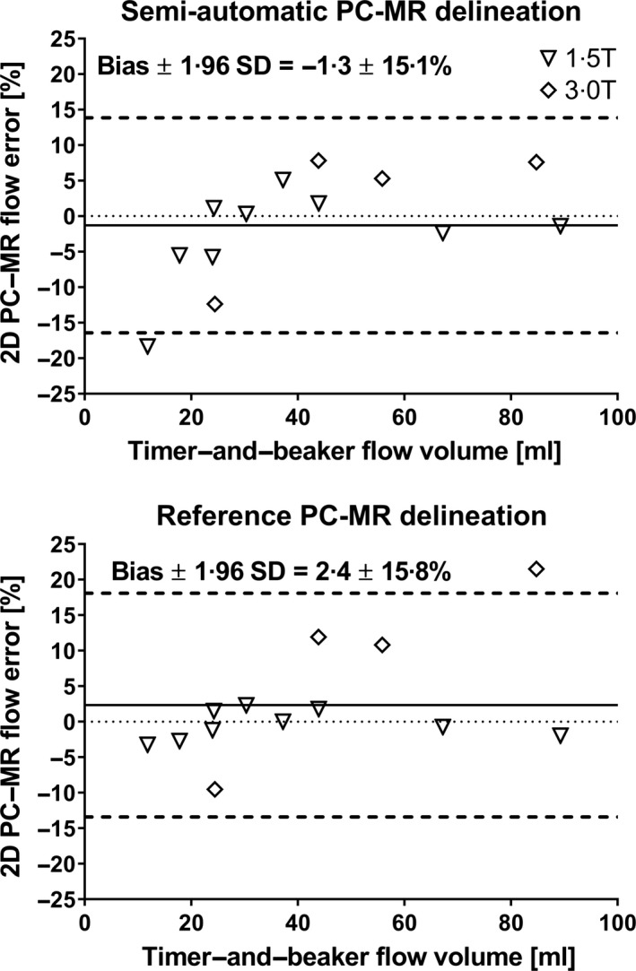
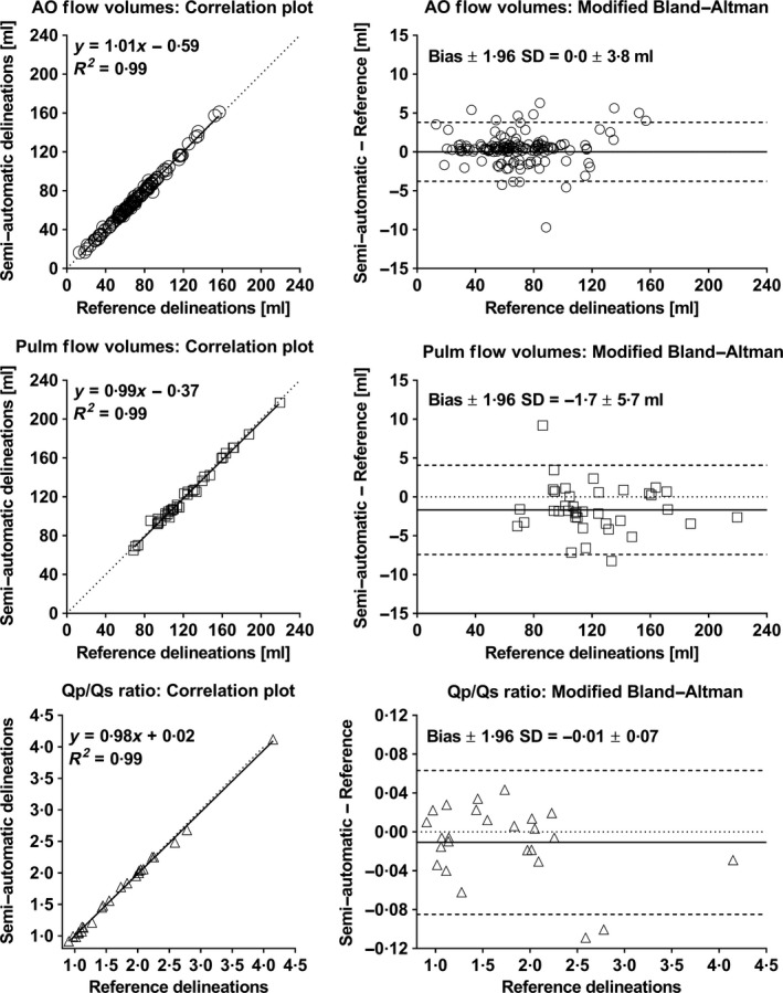
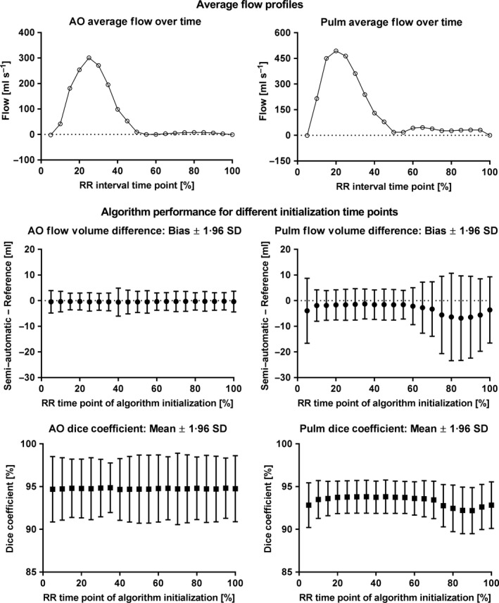
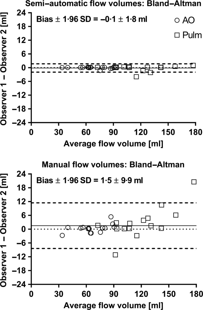
Blood flow measurements in the ascending aorta and pulmonary artery from phase-contrast magnetic resonance images require accurate time-resolved vessel segmentation over the cardiac cycle. Current semi-automatic segmentation methods often involve time-consuming manual correction, relying on user experience for accurate results. The purpose of this study was to develop a semi-automatic vessel segmentation algorithm with shape constraints based on manual vessel delineations for robust segmentation of the ascending aorta and pulmonary artery, to evaluate the proposed method in healthy volunteers and patients with heart failure and congenital heart disease, to validate the method in a pulsatile flow phantom experiment, and to make the method freely available for research purposes. Algorithm shape constraints were extracted from manual reference delineations of the ascending aorta (n = 20) and pulmonary artery (n = 20) and were included in a semi-automatic segmentation method only requiring manual delineation in one image. Bias and variability (bias ± SD) for flow volume of the proposed algorithm versus manual reference delineations were 0·0 ± 1·9 ml in the ascending aorta (n = 151; seven healthy volunteers; 144 heart failure patients) and -1·7 ± 2·9 ml in the pulmonary artery (n = 40; 25 healthy volunteers; 15 patients with atrial septal defect). Interobserver bias and variability were lower (P = 0·008) for the proposed semi-automatic method (-0·1 ± 0·9 ml) compared to manual reference delineations (1·5 ± 5·1 ml). Phantom validation showed good agreement between the proposed method and timer-and-beaker flow volumes (0·4 ± 2·7 ml). In conclusion, the proposed semi-automatic vessel segmentation algorithm can be used for efficient analysis of flow and shunt volumes in the aorta and pulmonary artery.

摘要

通过相位对比磁共振图像测量升主动脉和肺动脉的血流,需要在心动周期内对血管进行准确的时间分辨分割。当前的半自动分割方法通常需要耗时的人工校正,依赖用户经验才能获得准确结果。本研究的目的是基于手动血管描绘开发一种具有形状约束的半自动血管分割算法,用于稳健分割升主动脉和肺动脉;在健康志愿者、心力衰竭患者和先天性心脏病患者中评估所提出的方法;在脉动流体模实验中验证该方法;并使该方法免费用于研究目的。算法形状约束从升主动脉(n = 20)和肺动脉(n = 20)的手动参考描绘中提取,并包含在仅需在一幅图像中进行手动描绘的半自动分割方法中。在所提出算法与手动参考描绘之间,升主动脉(n = 151;7名健康志愿者;144名心力衰竭患者)的流量偏差和变异性(偏差±标准差)为0·0 ± 1·9 ml,肺动脉(n = 40;25名健康志愿者;15名房间隔缺损患者)为-1·7 ± 2·9 ml。与手动参考描绘(1·5 ± 5·1 ml)相比,所提出的半自动方法(-0·1 ± 0·9 ml)的观察者间偏差和变异性更低(P = 0·008)。体模验证表明所提出的方法与计时器和烧杯测量的流量之间具有良好的一致性(0·4 ± 2·7 ml)。总之,所提出的半自动血管分割算法可用于高效分析主动脉和肺动脉中的血流及分流体积。

https://cdn.ncbi.nlm.nih.gov/pmc/blobs/3ffd/6852024/080cb30f72f9/CPF-39-327-g006.jpg
https://cdn.ncbi.nlm.nih.gov/pmc/blobs/3ffd/6852024/ce8372ffcf29/CPF-39-327-g001.jpg
https://cdn.ncbi.nlm.nih.gov/pmc/blobs/3ffd/6852024/fcc1e0166610/CPF-39-327-g002.jpg
https://cdn.ncbi.nlm.nih.gov/pmc/blobs/3ffd/6852024/81ccc4107b08/CPF-39-327-g003.jpg
https://cdn.ncbi.nlm.nih.gov/pmc/blobs/3ffd/6852024/e3ba91eb39df/CPF-39-327-g004.jpg
https://cdn.ncbi.nlm.nih.gov/pmc/blobs/3ffd/6852024/b9c183f32ba8/CPF-39-327-g005.jpg
https://cdn.ncbi.nlm.nih.gov/pmc/blobs/3ffd/6852024/080cb30f72f9/CPF-39-327-g006.jpg

相似文献

1
A new vessel segmentation algorithm for robust blood flow quantification from two-dimensional phase-contrast magnetic resonance images.一种用于从二维相位对比磁共振图像中进行稳健血流定量的新型血管分割算法。
Clin Physiol Funct Imaging. 2019 Sep;39(5):327-338. doi: 10.1111/cpf.12582. Epub 2019 Jun 6.
2
Atlas-based analysis of 4D flow CMR: automated vessel segmentation and flow quantification.基于图谱的4D流心脏磁共振成像分析:自动血管分割与血流定量
J Cardiovasc Magn Reson. 2015 Oct 5;17:87. doi: 10.1186/s12968-015-0190-5.
3
In-vivo validation of interpolation-based phase offset correction in cardiovascular magnetic resonance flow quantification: a multi-vendor, multi-center study.基于内插的相位偏移校正在心血管磁共振流量定量中的体内验证:多供应商、多中心研究。
J Cardiovasc Magn Reson. 2019 May 20;21(1):30. doi: 10.1186/s12968-019-0538-3.
4
Intra- and inter-reader reproducibility of blood flow measurements on the ascending aorta and pulmonary artery using cardiac magnetic resonance.应用心脏磁共振评估升主动脉和肺动脉血流的测量的内、外重复性。
Radiol Med. 2017 Mar;122(3):179-185. doi: 10.1007/s11547-016-0706-6. Epub 2016 Nov 22.
5
The clinical impact of phase offset errors and different correction methods in cardiovascular magnetic resonance phase contrast imaging: a multi-scanner study.相位偏移误差及其在心血管磁共振相位对比成像中不同校正方法的临床影响:多台扫描仪研究。
J Cardiovasc Magn Reson. 2020 Sep 17;22(1):68. doi: 10.1186/s12968-020-00659-3.
6
Quantification of pulmonary/systemic shunt ratio by single-acquisition phase-contrast cardiovascular magnetic resonance.通过单次采集相位对比心血管磁共振定量肺/体分流率。
Echocardiography. 2019 Jun;36(6):1181-1190. doi: 10.1111/echo.14358. Epub 2019 May 13.
7
Automatic vessel segmentation using active contours in cine phase contrast flow measurements.在电影相位对比血流测量中使用活动轮廓进行血管自动分割。
J Magn Reson Imaging. 1999 Jul;10(1):41-51. doi: 10.1002/(sici)1522-2586(199907)10:1<41::aid-jmri6>3.0.co;2-j.
8
Machine learning derived segmentation of phase velocity encoded cardiovascular magnetic resonance for fully automated aortic flow quantification.机器学习分割相位速度编码心血管磁共振用于全自动主动脉流量定量分析。
J Cardiovasc Magn Reson. 2019 Jan 7;21(1):1. doi: 10.1186/s12968-018-0509-0.
9
Automatic detection of aorto-femoral vessel trajectory from whole-body computed tomography angiography data sets.从全身计算机断层扫描血管造影数据集自动检测主动脉-股动脉血管轨迹。
Int J Cardiovasc Imaging. 2016 Aug;32(8):1311-22. doi: 10.1007/s10554-016-0901-5. Epub 2016 May 21.
10
Improving the reproducibility of MR-derived left ventricular volume and function measurements with a semi-automatic threshold-based segmentation algorithm.使用基于半自动阈值分割算法提高心脏磁共振左心室容积和功能测量的可重复性。
Int J Cardiovasc Imaging. 2013 Mar;29(3):617-23. doi: 10.1007/s10554-012-0130-5. Epub 2012 Sep 29.

引用本文的文献

1
AI-powered automated model construction for patient-specific CFD simulations of aortic flows.用于主动脉血流患者特异性计算流体动力学模拟的人工智能驱动的自动模型构建。
Sci Adv. 2025 Sep 5;11(36):eadw2825. doi: 10.1126/sciadv.adw2825.
2
Cerebral Blood Flow under Pressure: Investigating Cerebrovascular Compliance with Phase-contrast Magnetic Resonance Imaging during Induced Hypertension.压力下的脑血流量:在诱导性高血压期间用相位对比磁共振成像研究脑血管顺应性
Anesthesiology. 2025 Oct 1;143(4):917-928. doi: 10.1097/ALN.0000000000005651. Epub 2025 Jul 11.
3
Plasma expansion and renal perfusion in critical COVID-19 with AKI: A prospective case control study.

本文引用的文献

1
Machine learning derived segmentation of phase velocity encoded cardiovascular magnetic resonance for fully automated aortic flow quantification.机器学习分割相位速度编码心血管磁共振用于全自动主动脉流量定量分析。
J Cardiovasc Magn Reson. 2019 Jan 7;21(1):1. doi: 10.1186/s12968-018-0509-0.
2
Changes in blood volume shunting in patients with atrial septal defects: assessment of heart function with cardiovascular magnetic resonance during dobutamine stress.房间隔缺损患者血液容量分流的变化:多巴酚丁胺负荷下心血管磁共振评估心功能。
Eur Heart J Cardiovasc Imaging. 2017 Oct 1;18(10):1145-1152. doi: 10.1093/ehjci/jew176.
3
新型冠状病毒肺炎合并急性肾损伤患者的血浆扩容与肾脏灌注:一项前瞻性病例对照研究
Acta Anaesthesiol Scand. 2025 Mar;69(3):e70004. doi: 10.1111/aas.70004.
4
Evaluation of Real-Time Cardiovascular Flow MRI Using Compressed Sensing in a Phantom and in Patients With Valvular Disease or Arrhythmia.使用压缩感知技术在体模以及患有瓣膜疾病或心律失常的患者中对实时心血管血流MRI进行评估。
J Magn Reson Imaging. 2025 Aug;62(2):417-429. doi: 10.1002/jmri.29702. Epub 2025 Jan 20.
5
Individualized cortical gyrification in neonates with congenital heart disease.先天性心脏病新生儿的个体化脑回形成
Brain Commun. 2024 Oct 7;6(5):fcae356. doi: 10.1093/braincomms/fcae356. eCollection 2024.
6
Subjects with carotid webs demonstrate pro-thrombotic hemodynamics compared to subjects with carotid atherosclerosis.与颈动脉粥样硬化患者相比,颈动脉膜患者表现出促血栓形成的血液动力学特征。
Sci Rep. 2024 May 2;14(1):10092. doi: 10.1038/s41598-024-60666-7.
7
Timing of Regadenoson-induced Peak Hyperemia and the Effects on Coronary Flow Reserve.雷加曲班诱导的峰值充血时间及其对冠状动脉血流储备的影响。
medRxiv. 2024 Jan 16:2024.01.15.23300449. doi: 10.1101/2024.01.15.23300449.
8
Uncertainty Quantification in the In Vivo Image-Based Estimation of Local Elastic Properties of Vascular Walls.基于体内图像的血管壁局部弹性特性估计中的不确定性量化
J Cardiovasc Dev Dis. 2023 Mar 4;10(3):109. doi: 10.3390/jcdd10030109.
9
Fetal growth restriction followed by very preterm birth is associated with smaller kidneys but preserved kidney function in adolescence.胎儿生长受限继而出现极早产与青春期时肾脏较小但肾功能正常有关。
Pediatr Nephrol. 2023 Jun;38(6):1855-1866. doi: 10.1007/s00467-022-05785-x. Epub 2022 Nov 21.
10
Fully-automated deep learning-based flow quantification of 2D CINE phase contrast MRI.基于全自动深度学习的二维 Cine 相位对比 MRI 流量定量分析。
Eur Radiol. 2023 Mar;33(3):1707-1718. doi: 10.1007/s00330-022-09179-3. Epub 2022 Oct 29.
Atlas-based analysis of 4D flow CMR: automated vessel segmentation and flow quantification.
基于图谱的4D流心脏磁共振成像分析:自动血管分割与血流定量
J Cardiovasc Magn Reson. 2015 Oct 5;17:87. doi: 10.1186/s12968-015-0190-5.
4
Validation and Development of a New Automatic Algorithm for Time-Resolved Segmentation of the Left Ventricle in Magnetic Resonance Imaging.磁共振成像中左心室时间分辨分割新自动算法的验证与开发
Biomed Res Int. 2015;2015:970357. doi: 10.1155/2015/970357. Epub 2015 Jun 21.
5
Independent validation of four-dimensional flow MR velocities and vortex ring volume using particle imaging velocimetry and planar laser-Induced fluorescence.使用粒子图像测速技术和平面激光诱导荧光对四维流动磁共振速度和涡环体积进行独立验证。
Magn Reson Med. 2016 Mar;75(3):1064-75. doi: 10.1002/mrm.25683. Epub 2015 May 2.
6
Fully automated tool to identify the aorta and compute flow using phase-contrast MRI: validation and application in a large population based study.使用相位对比磁共振成像识别主动脉并计算血流的全自动工具:在一项基于大样本人群的研究中的验证与应用
J Magn Reson Imaging. 2014 Jul;40(1):221-8. doi: 10.1002/jmri.24338. Epub 2013 Sep 30.
7
Cardiac output and cardiac index measured with cardiovascular magnetic resonance in healthy subjects, elite athletes and patients with congestive heart failure.健康受试者、精英运动员和充血性心力衰竭患者的心血管磁共振测量心输出量和心指数。
J Cardiovasc Magn Reson. 2012 Jul 28;14(1):51. doi: 10.1186/1532-429X-14-51.
8
Automated segmentation of the aorta from phase contrast MR images: validation against expert tracing in healthy volunteers and in patients with a dilated aorta.自动分割磁共振相位对比图像中的主动脉:在健康志愿者和扩张性主动脉患者中与专家追踪的验证。
J Magn Reson Imaging. 2010 Apr;31(4):881-8. doi: 10.1002/jmri.22124.
9
Flow measurement by cardiovascular magnetic resonance: a multi-centre multi-vendor study of background phase offset errors that can compromise the accuracy of derived regurgitant or shunt flow measurements.心血管磁共振流量测量:多中心多厂家背景相位偏移误差研究,该误差可能会影响反流或分流流量测量的准确性。
J Cardiovasc Magn Reson. 2010 Jan 14;12(1):5. doi: 10.1186/1532-429X-12-5.
10
Design and validation of Segment--freely available software for cardiovascular image analysis.设计和验证——用于心血管图像分析的免费软件段。
BMC Med Imaging. 2010 Jan 11;10:1. doi: 10.1186/1471-2342-10-1.