Nakamura Yoshifumi, Yamada Reiko, Kaneko Maki, Naota Hiroaki, Fujimura Yu, Tabata Masami, Kobayashi Kazuhiko, Tanaka Kyosuke
Department of Gastroenterology, Matsusaka Chuo General Hospital, Matsusaka, Mie, Japan.
Department of Gastroenterology and Hepatology, Mie University Hospital, 2-174 Edobashi, Tsu, Mie, 514-8507, Japan.
Clin J Gastroenterol. 2019 Dec;12(6):626-636. doi: 10.1007/s12328-019-00996-6. Epub 2019 May 27.
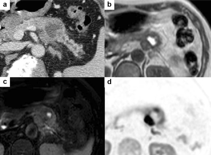
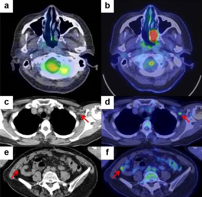
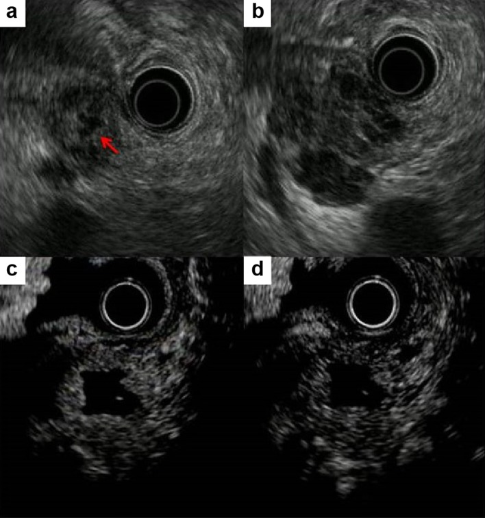
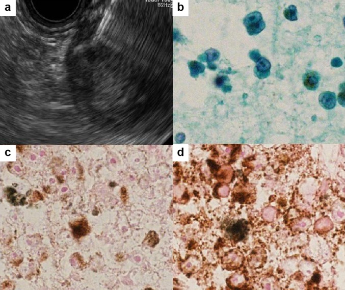
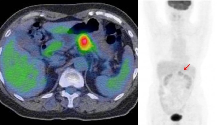
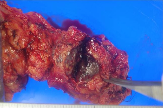
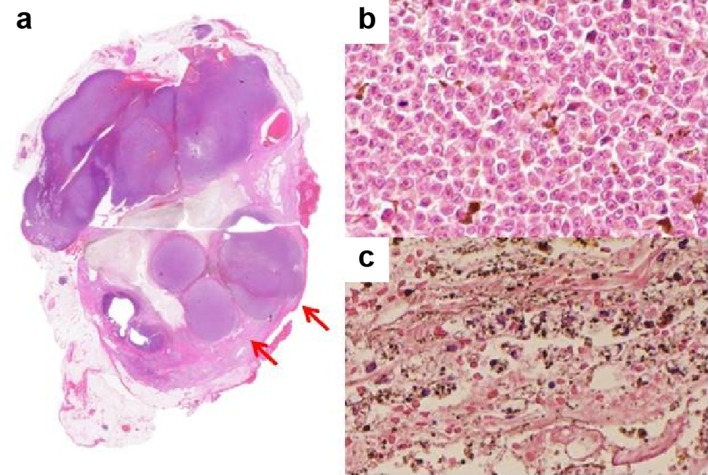
Isolated pancreatic metastasis from malignant melanoma is rare. Pancreatic metastasis is difficult to diagnose in patients with unknown primary malignant melanoma. Endoscopic ultrasound-guided fine-needle aspiration plays an important role in confirming the diagnosis. A 67-year-old woman was referred to our institution because of a mass in her pancreas. Computed tomography and magnetic resonance imaging revealed a 35-mm mass localized on the pancreatic tail, with low attenuation, surrounded by a high-attenuation rim. Endoscopic ultrasonography revealed a hypoechoic mass with central anechoic areas. Endoscopic ultrasound-guided fine-needle aspiration of the mass was performed, and the pathological diagnosis was malignant melanoma. Intense fluorodeoxyglucose uptake was observed in the pancreatic tail on positron emission tomography-computed tomography. No other malignant melanoma was found. Distal pancreatectomy was performed. Six months postoperatively, positron emission tomography-computed tomography revealed high uptake in the left nasal cavity, and biopsy revealed the mass to be a malignant melanoma, indicating that the primary site of the malignant melanoma was the left nasal cavity and that the pancreatic mass and peritoneal lesion were metastases. The patient had survived > 2 years after the distal pancreatectomy. Pancreatic resection of isolated pancreatic metastasis can possibly prolong survival; however, metastatic melanoma usually has poor prognosis.
恶性黑色素瘤孤立性胰腺转移罕见。对于原发性恶性黑色素瘤不明的患者,胰腺转移难以诊断。内镜超声引导下细针穿刺在确诊中起重要作用。一名67岁女性因胰腺肿物被转诊至我院。计算机断层扫描和磁共振成像显示胰腺尾部有一个35毫米的肿物,呈低密度,周围有高密度边缘。内镜超声显示一个低回声肿物,中央为无回声区。对该肿物进行了内镜超声引导下细针穿刺,病理诊断为恶性黑色素瘤。正电子发射断层扫描 - 计算机断层扫描显示胰腺尾部有强烈的氟脱氧葡萄糖摄取。未发现其他恶性黑色素瘤。实施了远端胰腺切除术。术后6个月,正电子发射断层扫描 - 计算机断层扫描显示左侧鼻腔有高摄取,活检显示肿物为恶性黑色素瘤,表明恶性黑色素瘤的原发部位是左侧鼻腔,胰腺肿物和腹膜病变为转移灶。该患者在远端胰腺切除术后存活超过2年。孤立性胰腺转移灶的胰腺切除可能延长生存期;然而,转移性黑色素瘤通常预后较差。