• 文献检索
  • 文档翻译
  • 深度研究
  • 学术资讯
  • Suppr Zotero 插件Zotero 插件
  • 邀请有礼
  • 套餐&价格
  • 历史记录
应用&插件
Suppr Zotero 插件Zotero 插件浏览器插件Mac 客户端Windows 客户端微信小程序
定价
高级版会员购买积分包购买API积分包
服务
文献检索文档翻译深度研究API 文档MCP 服务
关于我们
关于 Suppr公司介绍联系我们用户协议隐私条款
关注我们

Suppr 超能文献

核心技术专利:CN118964589B侵权必究
粤ICP备2023148730 号-1Suppr @ 2026

文献检索

告别复杂PubMed语法,用中文像聊天一样搜索,搜遍4000万医学文献。AI智能推荐,让科研检索更轻松。

立即免费搜索

文件翻译

保留排版,准确专业,支持PDF/Word/PPT等文件格式,支持 12+语言互译。

免费翻译文档

深度研究

AI帮你快速写综述,25分钟生成高质量综述,智能提取关键信息,辅助科研写作。

立即免费体验

动态磁敏感磁共振灌注成像在诊断放疗后脑转移复发中的应用:一项系统评价和荟萃分析

Dynamic susceptibility MR perfusion in diagnosing recurrent brain metastases after radiotherapy: A systematic review and meta-analysis.

作者信息

Kwee Robert M, Kwee Thomas C

机构信息

Department of Radiology, Zuyderland Medical Center, Heerlen/Sittard/Geleen, The Netherlands.

Department of Radiology, Nuclear Medicine and Molecular Imaging, University Medical Center Groningen, Groningen, The Netherlands.

出版信息

J Magn Reson Imaging. 2020 Feb;51(2):524-534. doi: 10.1002/jmri.26812. Epub 2019 May 31.

DOI:10.1002/jmri.26812
PMID:31150144
原文链接:https://pmc.ncbi.nlm.nih.gov/articles/PMC7004193/
Abstract

BACKGROUND

The diagnostic performance of dynamic susceptibility contrast (DSC) MR perfusion in discriminating treatment-related changes from recurrence in irradiated brain metastases is currently not completely clear.

PURPOSE

To systematically review the accuracy of DSC MR perfusion in diagnosing recurrent brain metastases after radiotherapy.

STUDY TYPE

Systematic review and meta-analysis.

SUBJECTS

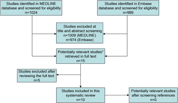
MEDLINE and Embase were searched for original studies investigating the accuracy of DSC MR perfusion in diagnosing recurrent brain metastases after radiotherapy. Ten studies, comprising a total of more than 271 metastases, were included.

FIELD STRENGTH/SEQUENCE: 1.5T or 3.0T, DSC MR perfusion.

ASSESSMENT

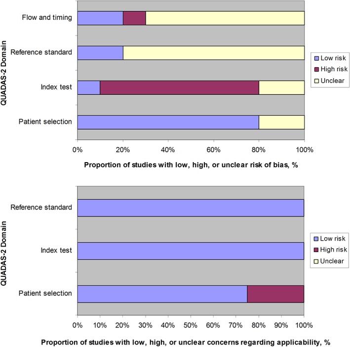
Quality assessment was performed according to the Quality Assessment of Diagnostic Accuracy Studies-2 tool.

STATISTICAL TESTS

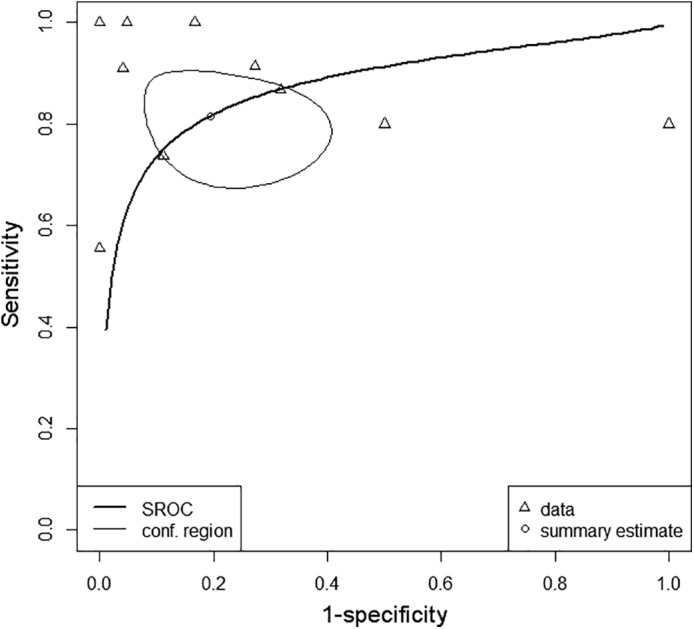
Sensitivity and specificity were pooled with a bivariate random-effects model. Heterogeneity was assessed by a chi-squared test. Potential sources for heterogeneity were explored by subgroup analyses.

RESULTS

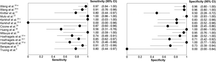
In seven studies the diagnostic criterion was not prespecified. In eight studies it was unclear whether the reference standard was interpreted blindly. In seven studies it was unclear whether DSC MR perfusion results influenced which reference standard was used. Pooled sensitivity and specificity were 81.6% (95% confidence interval [CI]: 70.6%, 89.1%) and 80.6% (95% CI: 64.2%, 90.6%), respectively. There was significant heterogeneity in both sensitivity (P = 0.005) and specificity (P < 0.001). There were no significant differences in relative diagnostic odds ratio according to publication year, country of origin, study size, and DSC MR perfusion interpretation method (visual analysis of cerebral blood volume [CBV] map vs. relative CBV measurement) (P > 0.2). Due to insufficiently detailed reporting, it was not possible to investigate the influence of primary tumor origin on accuracy.

DATA CONCLUSION

Our results suggest that the accuracy of DSC MR perfusion in diagnosing recurrent brain metastases after radiotherapy is fairly high. However, these findings should be interpreted with caution because of methodological quality concerns and heterogeneity between studies.

LEVEL OF EVIDENCE

3 Technical Efficacy: Stage 2 J. Magn. Reson. Imaging 2020;51:524-534.

摘要

背景

动态磁敏感对比增强(DSC)磁共振灌注成像在鉴别放疗后脑转移瘤的治疗相关改变与复发方面的诊断效能目前尚不完全明确。

目的

系统评价DSC磁共振灌注成像诊断放疗后脑转移瘤复发的准确性。

研究类型

系统评价和荟萃分析。

研究对象

检索MEDLINE和Embase数据库,查找关于DSC磁共振灌注成像诊断放疗后脑转移瘤复发准确性的原始研究。纳入10项研究,共涉及271个以上转移瘤。

场强/序列:1.5T或3.0T,DSC磁共振灌注成像。

评估

根据诊断准确性研究质量评估-2工具进行质量评估。

统计分析

采用双变量随机效应模型汇总敏感度和特异度。通过卡方检验评估异质性。通过亚组分析探索异质性的潜在来源。

结果

7项研究未预先设定诊断标准。8项研究未明确参考标准是否采用盲法解读。7项研究未明确DSC磁共振灌注成像结果是否影响参考标准的选择。汇总敏感度和特异度分别为81.6%(95%置信区间[CI]:70.6%,89.1%)和80.6%(95%CI:64.2%,90.6%)。敏感度(P = 0.005)和特异度(P < 0.001)均存在显著异质性。根据发表年份、研究来源国、研究规模和DSC磁共振灌注成像解读方法(脑血容量[CBV]图视觉分析与相对CBV测量),相对诊断比值比无显著差异(P > 0.2)。由于报告不够详细,无法研究原发肿瘤来源对准确性的影响。

数据结论

我们的结果表明,DSC磁共振灌注成像诊断放疗后脑转移瘤复发的准确性相当高。然而,由于方法学质量问题和研究间的异质性,这些结果应谨慎解读。

证据水平

3 技术效能:2级 《磁共振成像杂志》2020年;51:524 - 534。

https://cdn.ncbi.nlm.nih.gov/pmc/blobs/c012/7004193/fc8b724e48d0/JMRI-51-524-g007.jpg
https://cdn.ncbi.nlm.nih.gov/pmc/blobs/c012/7004193/6f32d39a51e8/JMRI-51-524-g001.jpg
https://cdn.ncbi.nlm.nih.gov/pmc/blobs/c012/7004193/038de79b3731/JMRI-51-524-g002.jpg
https://cdn.ncbi.nlm.nih.gov/pmc/blobs/c012/7004193/9ce720d09730/JMRI-51-524-g003.jpg
https://cdn.ncbi.nlm.nih.gov/pmc/blobs/c012/7004193/1383e9a22263/JMRI-51-524-g004.jpg
https://cdn.ncbi.nlm.nih.gov/pmc/blobs/c012/7004193/598ccc4b723f/JMRI-51-524-g005.jpg
https://cdn.ncbi.nlm.nih.gov/pmc/blobs/c012/7004193/b52461e99ce3/JMRI-51-524-g006.jpg
https://cdn.ncbi.nlm.nih.gov/pmc/blobs/c012/7004193/fc8b724e48d0/JMRI-51-524-g007.jpg

相似文献

1
Dynamic susceptibility MR perfusion in diagnosing recurrent brain metastases after radiotherapy: A systematic review and meta-analysis.动态磁敏感磁共振灌注成像在诊断放疗后脑转移复发中的应用:一项系统评价和荟萃分析
J Magn Reson Imaging. 2020 Feb;51(2):524-534. doi: 10.1002/jmri.26812. Epub 2019 May 31.
2
Prospective comparative diagnostic accuracy evaluation of dynamic contrast-enhanced (DCE) vs. dynamic susceptibility contrast (DSC) MR perfusion in differentiating tumor recurrence from radiation necrosis in treated high-grade gliomas.前瞻性对比诊断准确性评估:动态对比增强(DCE)与动态磁敏感对比(DSC)磁共振灌注成像在鉴别治疗后高级别脑胶质瘤肿瘤复发与放射性坏死中的作用。
J Magn Reson Imaging. 2019 Aug;50(2):573-582. doi: 10.1002/jmri.26621. Epub 2019 Jan 5.
3
Diagnostic Efficacy of Contrast-Enhanced MRI in Detecting Residual or Recurrent Hepatocellular Carcinoma After Transarterial Chemoembolization: A Systematic Review and Meta-analysis.经动脉化疗栓塞术后对比增强磁共振成像检测残余或复发性肝细胞癌的诊断效能:一项系统评价和荟萃分析
J Magn Reson Imaging. 2020 Oct;52(4):1019-1028. doi: 10.1002/jmri.26970. Epub 2019 Nov 1.
4
MRI as a diagnostic biomarker for differentiating primary central nervous system lymphoma from glioblastoma: A systematic review and meta-analysis.MRI 作为一种诊断生物标志物,用于区分原发性中枢神经系统淋巴瘤与胶质母细胞瘤:系统评价和荟萃分析。
J Magn Reson Imaging. 2019 Aug;50(2):560-572. doi: 10.1002/jmri.26602. Epub 2019 Jan 14.
5
Contrast-Enhanced Perfusion MR Imaging to Differentiate Between Recurrent/Residual Brain Neoplasms and Radiation Necrosis.对比增强灌注磁共振成像鉴别复发性/残留性脑肿瘤与放射性坏死
Asian Pac J Cancer Prev. 2018 Apr 25;19(4):941-948. doi: 10.22034/APJCP.2018.19.4.941.
6
Comparison of three different MR perfusion techniques and MR spectroscopy for multiparametric assessment in distinguishing recurrent high-grade gliomas from stable disease.比较三种不同的磁共振灌注技术和磁共振波谱在鉴别复发性高级别胶质瘤与稳定期疾病中的多参数评估。
Acad Radiol. 2013 Dec;20(12):1557-65. doi: 10.1016/j.acra.2013.09.003.
7
Comparison of Diffusion Tensor Imaging and Magnetic Resonance Perfusion Imaging in Differentiating Recurrent Brain Neoplasm From Radiation Necrosis.扩散张量成像与磁共振灌注成像在鉴别复发性脑肿瘤与放射性坏死中的比较
Acad Radiol. 2016 May;23(5):569-76. doi: 10.1016/j.acra.2015.11.015. Epub 2016 Feb 23.
8
Absolute CBV for the differentiation of recurrence and radionecrosis of brain metastases after gamma knife radiotherapy: a comparison with relative CBV.绝对脑血容量(CBV)用于鉴别脑转移瘤伽玛刀放射治疗后复发与放射性坏死:与相对 CBV 的比较。
Clin Radiol. 2018 Aug;73(8):758.e1-758.e7. doi: 10.1016/j.crad.2018.04.006. Epub 2018 Jun 18.
9
Utility of Percentage Signal Recovery and Baseline Signal in DSC-MRI Optimized for Relative CBV Measurement for Differentiating Glioblastoma, Lymphoma, Metastasis, and Meningioma.用于相对 CBV 测量的 DSC-MRI 的百分比信号恢复和基线信号的效用,可用于鉴别胶质母细胞瘤、淋巴瘤、转移瘤和脑膜瘤。
AJNR Am J Neuroradiol. 2019 Sep;40(9):1445-1450. doi: 10.3174/ajnr.A6153. Epub 2019 Aug 1.
10
Diagnostic performance of CT, gadoxetate disodium-enhanced MRI, and PET/CT for the diagnosis of colorectal liver metastasis: Systematic review and meta-analysis.CT、钆塞酸二钠增强 MRI 和 PET/CT 诊断结直肠癌肝转移的诊断性能:系统评价和荟萃分析。
J Magn Reson Imaging. 2018 May;47(5):1237-1250. doi: 10.1002/jmri.25852. Epub 2017 Sep 13.

引用本文的文献

1
Multidisciplinary management strategies for recurrent brain metastasis after prior radiotherapy: An overview.既往放疗后复发性脑转移瘤的多学科管理策略:综述
Neuro Oncol. 2025 Mar 7;27(3):597-615. doi: 10.1093/neuonc/noae220.
2
Gadolinium-Enhanced T2 FLAIR Is an Imaging Biomarker of Radiation Necrosis and Tumor Progression in Patients with Brain Metastases.钆增强T2液体衰减反转恢复序列是脑转移瘤患者放射性坏死和肿瘤进展的影像学生物标志物。
AJNR Am J Neuroradiol. 2025 Jan 8;46(1):129-135. doi: 10.3174/ajnr.A8431.
3
Prediction of radiologic outcome-optimized dose plans and post-treatment magnetic resonance images: A proof-of-concept study in breast cancer brain metastases treated with stereotactic radiosurgery.

本文引用的文献

1
A single-institution prospective evaluation of a neuro-oncology multidisciplinary team meeting.对一个神经肿瘤多学科团队会议进行的单机构前瞻性评估。
J Clin Neurosci. 2018 Oct;56:127-130. doi: 10.1016/j.jocn.2018.06.032. Epub 2018 Jun 29.
2
Absolute CBV for the differentiation of recurrence and radionecrosis of brain metastases after gamma knife radiotherapy: a comparison with relative CBV.绝对脑血容量(CBV)用于鉴别脑转移瘤伽玛刀放射治疗后复发与放射性坏死:与相对 CBV 的比较。
Clin Radiol. 2018 Aug;73(8):758.e1-758.e7. doi: 10.1016/j.crad.2018.04.006. Epub 2018 Jun 18.
3
Interval Change in Diffusion and Perfusion MRI Parameters for the Assessment of Pseudoprogression in Cerebral Metastases Treated With Stereotactic Radiation.
放射学结果预测——优化剂量计划与治疗后磁共振成像:立体定向放射外科治疗乳腺癌脑转移瘤的概念验证研究
Phys Imaging Radiat Oncol. 2024 Jun 24;31:100602. doi: 10.1016/j.phro.2024.100602. eCollection 2024 Jul.
4
Emerging therapeutics and evolving assessment criteria for intracranial metastases in patients with oncogene-driven non-small-cell lung cancer.驱动基因阳性的非小细胞肺癌颅内转移的新兴治疗方法和评估标准的演变。
Nat Rev Clin Oncol. 2023 Oct;20(10):716-732. doi: 10.1038/s41571-023-00808-4. Epub 2023 Aug 17.
5
Radiological Biomarkers for Brain Metastases Prognosis: Quantitative Magnetic Resonance Imaging (MRI) Modalities As Non-invasive Biomarkers for the Effect of Radiotherapy.用于脑转移预后的放射学生物标志物:定量磁共振成像(MRI)模态作为放疗效果的非侵入性生物标志物
Cureus. 2023 Apr 30;15(4):e38353. doi: 10.7759/cureus.38353. eCollection 2023 Apr.
6
Towards differentiation of brain tumor from radiation necrosis using multi-parametric MRI: Preliminary results at 4.7 T using rodent models.使用多参数 MRI 区分脑肿瘤与放射性坏死:在 4.7T 下使用啮齿动物模型的初步结果。
Magn Reson Imaging. 2022 Dec;94:144-150. doi: 10.1016/j.mri.2022.10.002. Epub 2022 Oct 6.
7
Selective Cell Size MRI Differentiates Brain Tumors from Radiation Necrosis.磁共振选择性细胞大小成像可区分脑肿瘤与放射性坏死。
Cancer Res. 2022 Oct 4;82(19):3603-3613. doi: 10.1158/0008-5472.CAN-21-2929.
8
Variability of relative cerebral blood volume measurements of recurrent glioma.复发性胶质瘤相对脑血容量测量的变异性
Ann Transl Med. 2019 Dec;7(Suppl 8):S260. doi: 10.21037/atm.2019.12.41.
磁共振弥散和灌注成像参数的时间变化在评估立体定向放疗治疗脑转移瘤假性进展中的应用。
AJR Am J Roentgenol. 2018 Jul;211(1):168-175. doi: 10.2214/AJR.17.18890. Epub 2018 Apr 30.
4
Observer variability of reference tissue selection for relativecerebral blood volume measurements in glioma patients.脑肿瘤患者相对脑血容量测量中参考组织选择的观察者变异性。
Eur Radiol. 2018 Sep;28(9):3902-3911. doi: 10.1007/s00330-018-5353-y. Epub 2018 Mar 23.
5
Dynamic susceptibility contrast (DSC) perfusion MRI in differential diagnosis between radionecrosis and neoangiogenesis in cerebral metastases using rCBV, rCBF and K2.动态对比磁共振灌注成像(DSC)在脑转移瘤放射性坏死与新生血管形成鉴别诊断中的应用:rCBV、rCBF 和 K2 的价值。
Radiol Med. 2018 Jul;123(7):545-552. doi: 10.1007/s11547-018-0866-7. Epub 2018 Mar 5.
6
Clinical applicability of and changes in perfusion MR imaging in brain metastases after stereotactic radiotherapy.立体定向放疗后脑转移瘤灌注磁共振成像的临床应用及变化。
J Neurooncol. 2018 May;138(1):133-139. doi: 10.1007/s11060-018-2779-7. Epub 2018 Feb 1.
7
Diagnostic Accuracy of Amino Acid and FDG-PET in Differentiating Brain Metastasis Recurrence from Radionecrosis after Radiotherapy: A Systematic Review and Meta-Analysis.氨基酸和 FDG-PET 鉴别放疗后脑转移复发与放射性坏死的诊断准确性:系统评价和荟萃分析。
AJNR Am J Neuroradiol. 2018 Feb;39(2):280-288. doi: 10.3174/ajnr.A5472. Epub 2017 Dec 14.
8
Two-year experience of multi-disciplinary team (MDT) outcomes for brain metastases in a tertiary neuro-oncology centre.三级神经肿瘤中心脑转移瘤多学科团队(MDT)治疗效果的两年经验
Br J Neurosurg. 2018 Feb;32(1):53-60. doi: 10.1080/02688697.2017.1368449. Epub 2017 Aug 24.
9
Incidence and prognosis of patients with brain metastases at diagnosis of systemic malignancy: a population-based study.初诊系统性恶性肿瘤患者脑转移的发生率和预后:一项基于人群的研究。
Neuro Oncol. 2017 Oct 19;19(11):1511-1521. doi: 10.1093/neuonc/nox077.
10
Follow-up results of brain metastasis patients undergoing repeat Gamma Knife radiosurgery.行重复伽玛刀放射外科治疗的脑转移瘤患者的随访结果。
J Neurosurg. 2016 Dec;125(Suppl 1):2-10. doi: 10.3171/2016.6.GKS161404.