• 文献检索
  • 文档翻译
  • 深度研究
  • 学术资讯
  • Suppr Zotero 插件Zotero 插件
  • 邀请有礼
  • 套餐&价格
  • 历史记录
应用&插件
Suppr Zotero 插件Zotero 插件浏览器插件Mac 客户端Windows 客户端微信小程序
定价
高级版会员购买积分包购买API积分包
服务
文献检索文档翻译深度研究API 文档MCP 服务
关于我们
关于 Suppr公司介绍联系我们用户协议隐私条款
关注我们

Suppr 超能文献

核心技术专利:CN118964589B侵权必究
粤ICP备2023148730 号-1Suppr @ 2026

文献检索

告别复杂PubMed语法,用中文像聊天一样搜索,搜遍4000万医学文献。AI智能推荐,让科研检索更轻松。

立即免费搜索

文件翻译

保留排版,准确专业,支持PDF/Word/PPT等文件格式,支持 12+语言互译。

免费翻译文档

深度研究

AI帮你快速写综述,25分钟生成高质量综述,智能提取关键信息,辅助科研写作。

立即免费体验

磁共振选择性细胞大小成像可区分脑肿瘤与放射性坏死。

Selective Cell Size MRI Differentiates Brain Tumors from Radiation Necrosis.

机构信息

Vanderbilt University Institute of Imaging Science, Vanderbilt University Medical Center, Nashville, Tennessee.

Chemical and Physical Biology Program, Vanderbilt University, Nashville, Tennessee.

出版信息

Cancer Res. 2022 Oct 4;82(19):3603-3613. doi: 10.1158/0008-5472.CAN-21-2929.

DOI:10.1158/0008-5472.CAN-21-2929
PMID:35877201
原文链接:https://pmc.ncbi.nlm.nih.gov/articles/PMC9532360/
Abstract

UNLABELLED

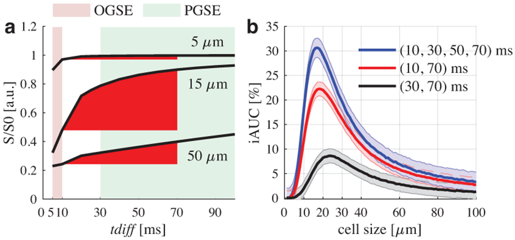
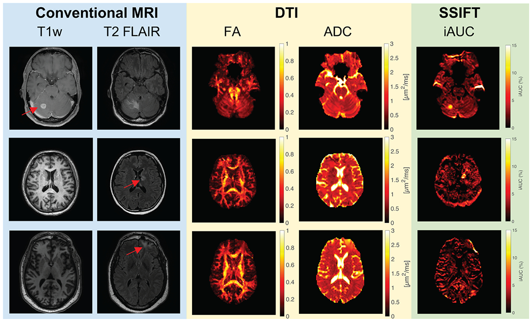
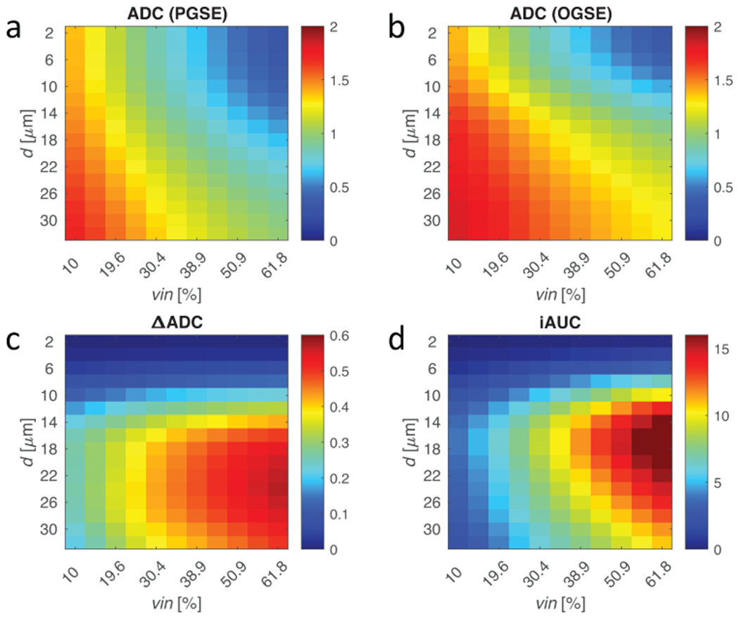
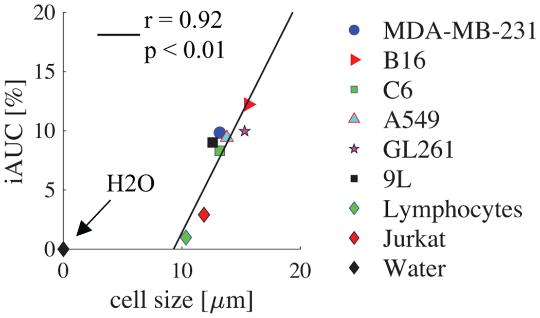
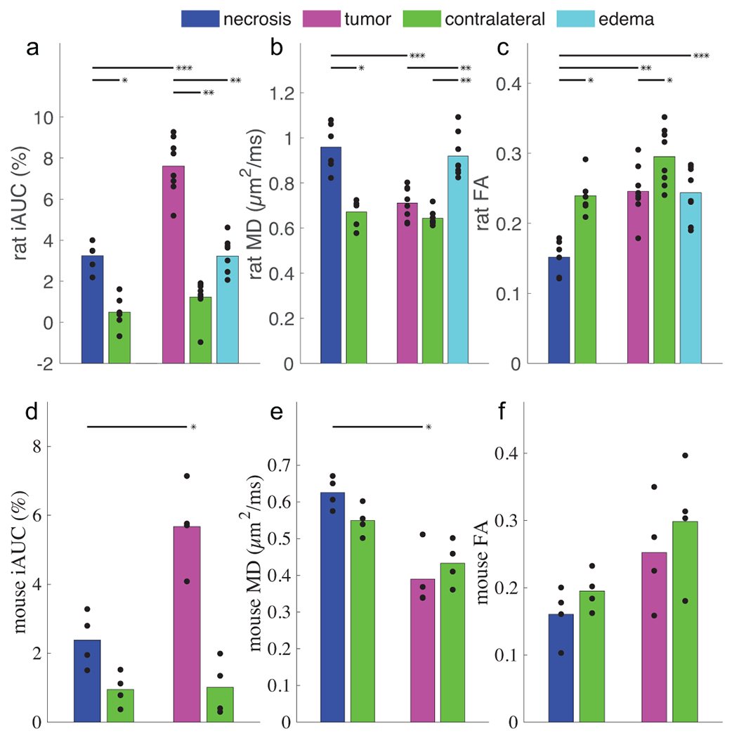
Brain metastasis is a common characteristic of late-stage lung cancers. High doses of targeted radiotherapy can control tumor growth in the brain but can also result in radiotherapy-induced necrosis. Current methods are limited for distinguishing whether new parenchymal lesions following radiotherapy are recurrent tumors or radiotherapy-induced necrosis, but the clinical management of these two classes of lesions differs significantly. Here, we developed, validated, and evaluated a new MRI technique termed selective size imaging using filters via diffusion times (SSIFT) to differentiate brain tumors from radiotherapy necrosis in the brain. This approach generates a signal filter that leverages diffusion time dependence to establish a cell size-weighted map. Computer simulations in silico, cultured cancer cells in vitro, and animals with brain tumors in vivo were used to comprehensively validate the specificity of SSIFT for detecting typical large cancer cells and the ability to differentiate brain tumors from radiotherapy necrosis. SSIFT was also implemented in patients with metastatic brain cancer and radiotherapy necrosis. SSIFT showed high correlation with mean cell sizes in the relevant range of less than 20 μm. The specificity of SSIFT for brain tumors and reduced contrast in other brain etiologies allowed SSIFT to differentiate brain tumors from peritumoral edema and radiotherapy necrosis. In conclusion, this new, cell size-based MRI method provides a unique contrast to differentiate brain tumors from other pathologies in the brain.

SIGNIFICANCE

This work introduces and provides preclinical validation of a new diffusion MRI method that exploits intrinsic differences in cell sizes to distinguish brain tumors and radiotherapy necrosis.

摘要

未加标签

脑转移是晚期肺癌的一个常见特征。高剂量的靶向放疗可以控制脑部肿瘤的生长,但也会导致放疗引起的坏死。目前的方法在区分放疗后新的实质病变是复发性肿瘤还是放疗引起的坏死方面存在局限性,但这两种病变的临床处理有很大的不同。在这里,我们开发、验证和评估了一种新的 MRI 技术,称为通过扩散时间使用滤波器的选择性大小成像(SSIFT),以区分脑部的肿瘤和放疗引起的坏死。这种方法生成一个信号滤波器,利用扩散时间依赖性来建立一个细胞大小加权图。在计算机模拟、体外培养的癌细胞和体内脑瘤动物中进行了全面验证,证明了 SSIFT 具有检测典型大癌细胞的特异性,以及区分脑瘤和放疗坏死的能力。SSIFT 还在转移性脑癌和放疗坏死的患者中得到了实施。SSIFT 与相关范围内小于 20μm 的平均细胞大小高度相关。SSIFT 对脑瘤的特异性和其他脑部病因对比度的降低,使 SSIFT 能够区分脑瘤与肿瘤周围水肿和放疗坏死。总之,这种新的基于细胞大小的 MRI 方法为区分脑部的肿瘤和其他病变提供了独特的对比。

意义

这项工作介绍并提供了一种新的扩散 MRI 方法的临床前验证,该方法利用细胞大小的内在差异来区分脑瘤和放疗引起的坏死。

https://cdn.ncbi.nlm.nih.gov/pmc/blobs/1fc6/9532360/290ebf0622c6/nihms-1827204-f0006.jpg
https://cdn.ncbi.nlm.nih.gov/pmc/blobs/1fc6/9532360/76006506c751/nihms-1827204-f0001.jpg
https://cdn.ncbi.nlm.nih.gov/pmc/blobs/1fc6/9532360/ecc49117344d/nihms-1827204-f0002.jpg
https://cdn.ncbi.nlm.nih.gov/pmc/blobs/1fc6/9532360/b70ba4f68c03/nihms-1827204-f0003.jpg
https://cdn.ncbi.nlm.nih.gov/pmc/blobs/1fc6/9532360/de9cf4c9f044/nihms-1827204-f0004.jpg
https://cdn.ncbi.nlm.nih.gov/pmc/blobs/1fc6/9532360/7ad5e44f282a/nihms-1827204-f0005.jpg
https://cdn.ncbi.nlm.nih.gov/pmc/blobs/1fc6/9532360/290ebf0622c6/nihms-1827204-f0006.jpg

相似文献

1
Selective Cell Size MRI Differentiates Brain Tumors from Radiation Necrosis.磁共振选择性细胞大小成像可区分脑肿瘤与放射性坏死。
Cancer Res. 2022 Oct 4;82(19):3603-3613. doi: 10.1158/0008-5472.CAN-21-2929.
2
Comparison of Diffusion Tensor Imaging and Magnetic Resonance Perfusion Imaging in Differentiating Recurrent Brain Neoplasm From Radiation Necrosis.扩散张量成像与磁共振灌注成像在鉴别复发性脑肿瘤与放射性坏死中的比较
Acad Radiol. 2016 May;23(5):569-76. doi: 10.1016/j.acra.2015.11.015. Epub 2016 Feb 23.
3
Contrast-Enhanced Perfusion MR Imaging to Differentiate Between Recurrent/Residual Brain Neoplasms and Radiation Necrosis.对比增强灌注磁共振成像鉴别复发性/残留性脑肿瘤与放射性坏死
Asian Pac J Cancer Prev. 2018 Apr 25;19(4):941-948. doi: 10.22034/APJCP.2018.19.4.941.
4
Differentiation of Recurrent Glioblastoma from Delayed Radiation Necrosis by Using Voxel-based Multiparametric Analysis of MR Imaging Data.基于体素的磁共振成像数据分析对复发性脑胶质母细胞瘤与迟发性放射性坏死的鉴别诊断。
Radiology. 2017 Oct;285(1):206-213. doi: 10.1148/radiol.2017161588. Epub 2017 May 23.
5
Differentiating Radiation Necrosis from Brain Tumor Using Hyperpolarized Carbon-13 MR Metabolic Imaging.使用 13C 磁共振代谢成像鉴别放射性坏死与脑肿瘤。
Mol Imaging Biol. 2021 Jun;23(3):417-426. doi: 10.1007/s11307-020-01574-w. Epub 2021 Jan 13.
6
Differentiation between recurrent tumor and radiation necrosis in a child with anaplastic ependymoma after chemotherapy and radiation therapy.化疗和放疗后患有间变性室管膜瘤的儿童复发性肿瘤与放射性坏死的鉴别。
Strahlenther Onkol. 2003 Dec;179(12):819-22. doi: 10.1007/s00066-003-1141-x.
7
Towards differentiation of brain tumor from radiation necrosis using multi-parametric MRI: Preliminary results at 4.7 T using rodent models.使用多参数 MRI 区分脑肿瘤与放射性坏死:在 4.7T 下使用啮齿动物模型的初步结果。
Magn Reson Imaging. 2022 Dec;94:144-150. doi: 10.1016/j.mri.2022.10.002. Epub 2022 Oct 6.
8
Differentiating radiation necrosis from tumor progression in brain metastases treated with stereotactic radiotherapy: utility of intravoxel incoherent motion perfusion MRI and correlation with histopathology.在立体定向放疗治疗脑转移瘤中,从肿瘤进展中鉴别放射性坏死:应用体素内不相干运动灌注 MRI 的效用及其与组织病理学的相关性。
J Neurooncol. 2017 Sep;134(2):433-441. doi: 10.1007/s11060-017-2545-2. Epub 2017 Jul 3.
9
Disorder in Pixel-Level Edge Directions on T1WI Is Associated with the Degree of Radiation Necrosis in Primary and Metastatic Brain Tumors: Preliminary Findings.T1WI 上像素级边缘方向紊乱与原发性和转移性脑肿瘤的放射坏死程度相关:初步研究结果。
AJNR Am J Neuroradiol. 2019 Mar;40(3):412-417. doi: 10.3174/ajnr.A5958. Epub 2019 Feb 7.
10
Leveraging radiomics and machine learning to differentiate radiation necrosis from recurrence in patients with brain metastases.利用放射组学和机器学习区分脑转移瘤患者的放射性坏死与复发。
J Neurooncol. 2024 Jun;168(2):307-316. doi: 10.1007/s11060-024-04669-4. Epub 2024 Apr 30.

引用本文的文献

1
The need for evidence-based, outcome-focused medical imaging research for cancer management.癌症管理中基于证据、以结果为导向的医学影像研究的必要性。
Npj Imaging. 2025 Feb 3;3(1):6. doi: 10.1038/s44303-024-00067-7.
2
Engineering clinical translation of OGSE diffusion MRI.OGSE扩散磁共振成像的工程临床翻译
Magn Reson Med. 2025 Sep;94(3):913-936. doi: 10.1002/mrm.30510. Epub 2025 May 7.
3
Assessing tumor microstructure with time-dependent diffusion imaging: Considerations and feasibility on clinical MRI and MRI-Linac.利用时间依赖性扩散成像评估肿瘤微观结构:临床MRI和MRI直线加速器的考量与可行性

本文引用的文献

1
Time-Dependent Diffusion MRI for Quantitative Microstructural Mapping of Prostate Cancer.基于时间的扩散 MRI 用于前列腺癌的定量微观结构成像。
Radiology. 2022 Jun;303(3):578-587. doi: 10.1148/radiol.211180. Epub 2022 Mar 8.
2
MR cell size imaging with temporal diffusion spectroscopy.采用时扩散光谱法的磁共振细胞大小成像。
Magn Reson Imaging. 2021 Apr;77:109-123. doi: 10.1016/j.mri.2020.12.010. Epub 2020 Dec 16.
3
Optimization and numerical evaluation of multi-compartment diffusion MRI using the spherical mean technique for practical multiple sclerosis imaging.
Med Phys. 2025 Jan;52(1):346-361. doi: 10.1002/mp.17453. Epub 2024 Oct 10.
4
Multiparametric MRI for characterization of the tumour microenvironment.多参数 MRI 用于肿瘤微环境的特征分析。
Nat Rev Clin Oncol. 2024 Jun;21(6):428-448. doi: 10.1038/s41571-024-00891-1. Epub 2024 Apr 19.
5
Advances in the application of neuroinflammatory molecular imaging in brain malignancies.神经炎症分子成像在脑恶性肿瘤中的应用进展。
Front Immunol. 2023 Jul 18;14:1211900. doi: 10.3389/fimmu.2023.1211900. eCollection 2023.
6
CEST2022: Amide proton transfer-weighted MRI improves the diagnostic performance of multiparametric non-contrast-enhanced MRI techniques in patients with post-treatment high-grade gliomas.CEST2022:酰胺质子转移加权 MRI 提高了治疗后高级别脑胶质瘤患者多参数非对比增强 MRI 技术的诊断性能。
Magn Reson Imaging. 2023 Oct;102:222-228. doi: 10.1016/j.mri.2023.06.003. Epub 2023 Jun 14.
7
Temporal Diffusion Ratio (TDR) for imaging restricted diffusion: Optimisation and pre-clinical demonstration.用于成像受限扩散的时间扩散比(TDR):优化和临床前验证。
Neuroimage. 2023 Apr 1;269:119930. doi: 10.1016/j.neuroimage.2023.119930. Epub 2023 Feb 5.
8
Towards differentiation of brain tumor from radiation necrosis using multi-parametric MRI: Preliminary results at 4.7 T using rodent models.使用多参数 MRI 区分脑肿瘤与放射性坏死:在 4.7T 下使用啮齿动物模型的初步结果。
Magn Reson Imaging. 2022 Dec;94:144-150. doi: 10.1016/j.mri.2022.10.002. Epub 2022 Oct 6.
使用球平均技术对多室扩散 MRI 进行优化和数值评估,以用于实际的多发性硬化成像。
Magn Reson Imaging. 2020 Dec;74:56-63. doi: 10.1016/j.mri.2020.09.002. Epub 2020 Sep 6.
4
Feasibility of oscillating and pulsed gradient diffusion MRI to assess neonatal hypoxia-ischemia on clinical systems.在临床系统上评估新生儿缺氧缺血的震荡和脉冲梯度弥散 MRI 的可行性。
J Cereb Blood Flow Metab. 2021 Jun;41(6):1240-1250. doi: 10.1177/0271678X20944353. Epub 2020 Aug 18.
5
Noninvasive diffusion magnetic resonance imaging of brain tumour cell size for the early detection of therapeutic response.无创性扩散磁共振成像检测脑肿瘤细胞大小以早期发现治疗反应。
Sci Rep. 2020 Jun 8;10(1):9223. doi: 10.1038/s41598-020-65956-4.
6
Mapping hepatocyte size in vivo using temporal diffusion spectroscopy MRI.利用时扩散光谱 MRI 对体内肝细胞大小进行成像。
Magn Reson Med. 2020 Nov;84(5):2671-2683. doi: 10.1002/mrm.28299. Epub 2020 Apr 25.
7
Magnetic resonance imaging of mean cell size in human breast tumors.人类乳腺肿瘤平均细胞大小的磁共振成像
Magn Reson Med. 2020 Jun;83(6):2002-2014. doi: 10.1002/mrm.28056. Epub 2019 Nov 25.
8
MRtrix3: A fast, flexible and open software framework for medical image processing and visualisation.MRtrix3:一个用于医学图像处理和可视化的快速、灵活、开放的软件框架。
Neuroimage. 2019 Nov 15;202:116137. doi: 10.1016/j.neuroimage.2019.116137. Epub 2019 Aug 29.
9
Dynamic susceptibility MR perfusion in diagnosing recurrent brain metastases after radiotherapy: A systematic review and meta-analysis.动态磁敏感磁共振灌注成像在诊断放疗后脑转移复发中的应用:一项系统评价和荟萃分析
J Magn Reson Imaging. 2020 Feb;51(2):524-534. doi: 10.1002/jmri.26812. Epub 2019 May 31.
10
Non-invasive assessment of glioma microstructure using VERDICT MRI: correlation with histology.使用 VERDICT MRI 无创评估脑胶质瘤的微观结构:与组织学的相关性。
Eur Radiol. 2019 Oct;29(10):5559-5566. doi: 10.1007/s00330-019-6011-8. Epub 2019 Mar 19.