• 文献检索
  • 文档翻译
  • 深度研究
  • 学术资讯
  • Suppr Zotero 插件Zotero 插件
  • 邀请有礼
  • 套餐&价格
  • 历史记录
应用&插件
Suppr Zotero 插件Zotero 插件浏览器插件Mac 客户端Windows 客户端微信小程序
定价
高级版会员购买积分包购买API积分包
服务
文献检索文档翻译深度研究API 文档MCP 服务
关于我们
关于 Suppr公司介绍联系我们用户协议隐私条款
关注我们

Suppr 超能文献

核心技术专利:CN118964589B侵权必究
粤ICP备2023148730 号-1Suppr @ 2026

文献检索

告别复杂PubMed语法,用中文像聊天一样搜索,搜遍4000万医学文献。AI智能推荐,让科研检索更轻松。

立即免费搜索

文件翻译

保留排版,准确专业,支持PDF/Word/PPT等文件格式,支持 12+语言互译。

免费翻译文档

深度研究

AI帮你快速写综述,25分钟生成高质量综述,智能提取关键信息,辅助科研写作。

立即免费体验

基于识别成像引导的原子力显微镜力谱:一种用于绘制和研究低横向密度下相互作用位点的新模式。

AFM-Based Force Spectroscopy Guided by Recognition Imaging: A New Mode for Mapping and Studying Interaction Sites at Low Lateral Density.

作者信息

Koehler Melanie, Fis Anny, Gruber Hermann J, Hinterdorfer Peter

机构信息

Institute of Biophysics, Johannes Kepler University, 4020 Linz, Austria.

Louvain Institute of Biomolecular Science and Technology, Université Catholique de Louvain, 1348 Louvain-La-Neuve, Belgium.

出版信息

Methods Protoc. 2019 Jan 8;2(1):6. doi: 10.3390/mps2010006.

DOI:10.3390/mps2010006
PMID:31164590
原文链接:https://pmc.ncbi.nlm.nih.gov/articles/PMC6481044/
Abstract

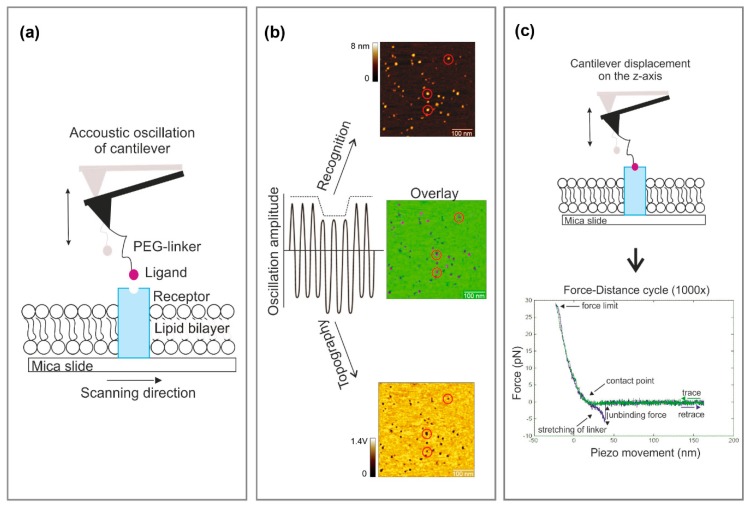
Ligand binding to receptors is one of the most important regulatory elements in biology as it is the initiating step in signaling pathways and cascades. Thus, precisely localizing binding sites and measuring interaction forces between cognate receptor-ligand pairs leads to new insights into the molecular recognition involved in these processes. Here we present a detailed protocol about applying a technique, which combines atomic force microscopy (AFM)-based recognition imaging and force spectroscopy for studying the interaction between (membrane) receptors and ligands on the single molecule level. This method allows for the selection of a single receptor molecule reconstituted into a supported lipid membrane at low density, with the subsequent quantification of the receptor-ligand unbinding force. Based on AFM tapping mode, a cantilever tip carrying a ligand molecule is oscillated across a membrane. Topography and recognition images of reconstituted receptors are recorded simultaneously by analyzing the downward and upward parts of the oscillation, respectively. Functional receptor molecules are selected from the recognition image with nanometer resolution before the AFM is switched to the force spectroscopy mode, using positional feedback control. The combined mode allows for dynamic force probing on different pre-selected molecules. This strategy results in higher throughput when compared with force mapping. Applied to two different receptor-ligand pairs, we validated the presented new mode.

摘要

配体与受体的结合是生物学中最重要的调节元件之一,因为它是信号通路和级联反应的起始步骤。因此,精确地定位结合位点并测量同源受体 - 配体对之间的相互作用力,能够让我们对这些过程中涉及的分子识别有新的认识。在此,我们展示了一个详细的实验方案,该方案应用了一种技术,即将基于原子力显微镜(AFM)的识别成像和力谱技术相结合,用于在单分子水平上研究(膜)受体与配体之间的相互作用。这种方法允许选择低密度重构于支撑脂质膜中的单个受体分子,随后对受体 - 配体解离力进行定量分析。基于AFM轻敲模式,一个携带配体分子的悬臂尖端在膜上振荡。通过分别分析振荡的向下和向上部分,同时记录重构受体的形貌和识别图像。在AFM切换到力谱模式之前,利用位置反馈控制从具有纳米分辨率的识别图像中选择功能性受体分子。这种组合模式允许对不同的预选分子进行动态力探测。与力映射相比,该策略具有更高的通量。应用于两对不同的受体 - 配体对,我们验证了所展示的新模式。

https://cdn.ncbi.nlm.nih.gov/pmc/blobs/5c78/6481044/a43384fd1669/mps-02-00006-g004.jpg
https://cdn.ncbi.nlm.nih.gov/pmc/blobs/5c78/6481044/85c616460609/mps-02-00006-g001.jpg
https://cdn.ncbi.nlm.nih.gov/pmc/blobs/5c78/6481044/5ecf5a92ff79/mps-02-00006-g002.jpg
https://cdn.ncbi.nlm.nih.gov/pmc/blobs/5c78/6481044/c0ea8a5de554/mps-02-00006-g003.jpg
https://cdn.ncbi.nlm.nih.gov/pmc/blobs/5c78/6481044/a43384fd1669/mps-02-00006-g004.jpg

相似文献

1
AFM-Based Force Spectroscopy Guided by Recognition Imaging: A New Mode for Mapping and Studying Interaction Sites at Low Lateral Density.基于识别成像引导的原子力显微镜力谱:一种用于绘制和研究低横向密度下相互作用位点的新模式。
Methods Protoc. 2019 Jan 8;2(1):6. doi: 10.3390/mps2010006.
2
Combined Recognition Imaging and Force Spectroscopy: A New Mode for Mapping and Studying Interaction Sites at Low Lateral Density.联合识别成像与力谱:一种用于在低横向密度下绘制和研究相互作用位点的新模式。
Sci Adv Mater. 2017 Jan 1;9(1):128-134. doi: 10.1166/sam.2017.3066.
3
Recent Progress in Molecular Recognition Imaging Using Atomic Force Microscopy.使用原子力显微镜进行分子识别成像的最新进展
Acc Chem Res. 2016 Mar 15;49(3):503-10. doi: 10.1021/acs.accounts.5b00533. Epub 2016 Mar 2.
4
Size Measurement of Nanoparticles Using Atomic Force Microscopy: Version 1.1使用原子力显微镜测量纳米颗粒的尺寸:版本1.1
5
Detection and localization of single molecular recognition events using atomic force microscopy.使用原子力显微镜检测和定位单分子识别事件。
Nat Methods. 2006 May;3(5):347-55. doi: 10.1038/nmeth871.
6
Simultaneous height and adhesion imaging of antibody-antigen interactions by atomic force microscopy.通过原子力显微镜对抗体-抗原相互作用进行高度和粘附力同步成像。
Biophys J. 1998 Nov;75(5):2220-8. doi: 10.1016/S0006-3495(98)77666-0.
7
Studying integrin-mediated cell adhesion at the single-molecule level using AFM force spectroscopy.使用原子力显微镜力谱在单分子水平研究整合素介导的细胞黏附。
Sci STKE. 2007 Oct 2;2007(406):pl5. doi: 10.1126/stke.4062007pl5.
8
Probing and mapping the binding sites on streptavidin imprinted polymer surface.探测并绘制链霉亲和素印迹聚合物表面的结合位点。
Mater Sci Eng C Mater Biol Appl. 2014 Oct;43:214-20. doi: 10.1016/j.msec.2014.07.018. Epub 2014 Jul 10.
9
Antibody recognition imaging by force microscopy.通过力显微镜进行抗体识别成像。
Nat Biotechnol. 1999 Sep;17(9):901-5. doi: 10.1038/12898.
10
Ligand-Receptor Binding on Cell Membrane: Dynamic Force Spectroscopy Applications.细胞膜上的配体-受体结合:动态力谱学应用
Methods Mol Biol. 2019;1886:153-162. doi: 10.1007/978-1-4939-8894-5_8.

引用本文的文献

1
Scanning Probe Microscopies: Imaging and Biomechanics in Reproductive Medicine Research.扫描探针显微镜技术:生殖医学研究中的成像与生物力学
Int J Mol Sci. 2021 Apr 7;22(8):3823. doi: 10.3390/ijms22083823.
2
Single-Molecule Force Spectroscopy Reveals Adhesion-by-Demand in Statherin at the Protein-Hydroxyapatite Interface.单分子力谱揭示了在蛋白质-羟磷灰石界面处的按需黏附现象。
Langmuir. 2020 Nov 10;36(44):13292-13300. doi: 10.1021/acs.langmuir.0c02325. Epub 2020 Oct 29.
3
Review: Cantilever-Based Sensors for High Speed Atomic Force Microscopy.

本文引用的文献

1
Combined Recognition Imaging and Force Spectroscopy: A New Mode for Mapping and Studying Interaction Sites at Low Lateral Density.联合识别成像与力谱:一种用于在低横向密度下绘制和研究相互作用位点的新模式。
Sci Adv Mater. 2017 Jan 1;9(1):128-134. doi: 10.1166/sam.2017.3066.
2
Inhibition of mitochondrial UCP1 and UCP3 by purine nucleotides and phosphate.嘌呤核苷酸和磷酸盐对线粒体 UCP1 和 UCP3 的抑制作用。
Biochim Biophys Acta Biomembr. 2018 Mar;1860(3):664-672. doi: 10.1016/j.bbamem.2017.12.001. Epub 2017 Dec 5.
3
The expression of UCP3 directly correlates to UCP1 abundance in brown adipose tissue.
综述:基于悬臂梁的高速原子力显微镜传感器。
Sensors (Basel). 2020 Aug 25;20(17):4784. doi: 10.3390/s20174784.
4
A new tool to determine the cellular metabolic landscape: nanotechnology to the study of Friedreich's ataxia.一种新的工具来确定细胞代谢特征:纳米技术在弗里德里希共济失调症研究中的应用。
Sci Rep. 2019 Dec 17;9(1):19282. doi: 10.1038/s41598-019-55799-z.
5
Mechanics of Brain Tissues Studied by Atomic Force Microscopy: A Perspective.原子力显微镜研究脑组织力学:综述
Front Neurosci. 2019 Jun 14;13:600. doi: 10.3389/fnins.2019.00600. eCollection 2019.
解偶联蛋白3(UCP3)的表达与棕色脂肪组织中解偶联蛋白1(UCP1)的丰度直接相关。
Biochim Biophys Acta. 2016 Jan;1857(1):72-78. doi: 10.1016/j.bbabio.2015.10.011. Epub 2015 Oct 27.
4
Ion conductivity of the bacterial translocation channel SecYEG engaged in translocation.参与转运的细菌转运通道SecYEG的离子电导率。
J Biol Chem. 2014 Aug 29;289(35):24611-6. doi: 10.1074/jbc.M114.588491. Epub 2014 Jul 11.
5
Comparison of three competing dynamic force spectroscopy models to study binding forces of amyloid-β (1-42).比较三种竞争的动态力谱模型以研究淀粉样β(1-42)的结合力。
Soft Matter. 2014 Mar 28;10(12):1924-30. doi: 10.1039/c3sm52257a.
6
The bacterial translocon SecYEG opens upon ribosome binding.细菌转运通道 SecYEG 在与核糖体结合时打开。
J Biol Chem. 2013 Jun 21;288(25):17941-6. doi: 10.1074/jbc.M113.477893. Epub 2013 May 3.
7
Dynamic structure of the translocon SecYEG in membrane: direct single molecule observations.膜中易位子 SecYEG 的动态结构:直接单分子观察。
J Biol Chem. 2013 Jun 7;288(23):16848-16854. doi: 10.1074/jbc.M113.471870. Epub 2013 Apr 22.
8
Mapping the nucleotide binding site of uncoupling protein 1 using atomic force microscopy.利用原子力显微镜绘制解偶联蛋白 1 的核苷酸结合位点。
J Am Chem Soc. 2013 Mar 6;135(9):3640-6. doi: 10.1021/ja312550k. Epub 2013 Feb 26.
9
Interpreting the widespread nonlinear force spectra of intermolecular bonds.解析分子间键的广泛非线性力谱。
Proc Natl Acad Sci U S A. 2012 Aug 21;109(34):13573-8. doi: 10.1073/pnas.1202946109. Epub 2012 Aug 6.
10
Glyphosate-induced stiffening of HaCaT keratinocytes, a Peak Force Tapping study on living cells.草甘膦诱导 HaCaT 角质形成细胞变硬:活细胞的峰力敲击研究。
J Struct Biol. 2012 Apr;178(1):1-7. doi: 10.1016/j.jsb.2012.02.007. Epub 2012 Feb 17.