Huang Qingling, Cao Xuan, Chai Xue, Wang Xiao, Xu Ligang, Xiao Chaoyong
Department of Radiology, Nanjing Medical University Affiliated Nanjing Brain Hospital, Nanjing, China.
Department of Mathematical Sciences, University of Cincinnati, Cincinnati, USA.
Medicine (Baltimore). 2019 Jun;98(23):e15972. doi: 10.1097/MD.0000000000015972.
This study aimed to evaluate the value of 3-dimensional pseudocontinuous arterial spin labeling (3D-pcASL) and susceptibility-weighted imaging (SWI) for the early disease-sensitive markers of conversion from amnestic MCI (aMCI) to Alzheimer disease (AD) in this process.
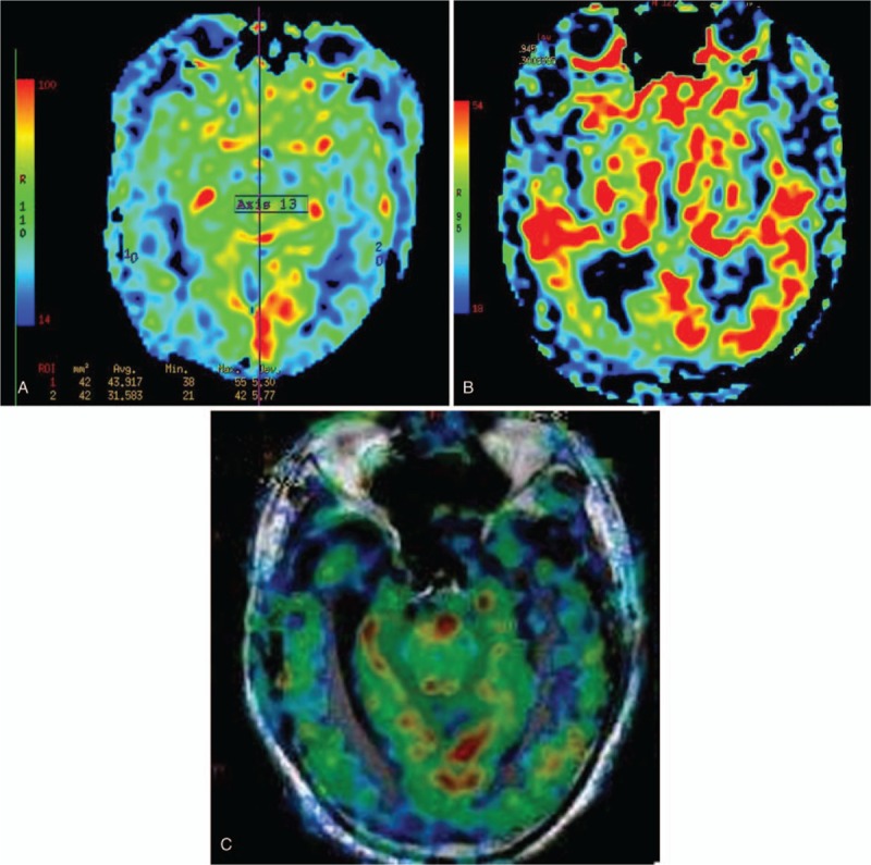
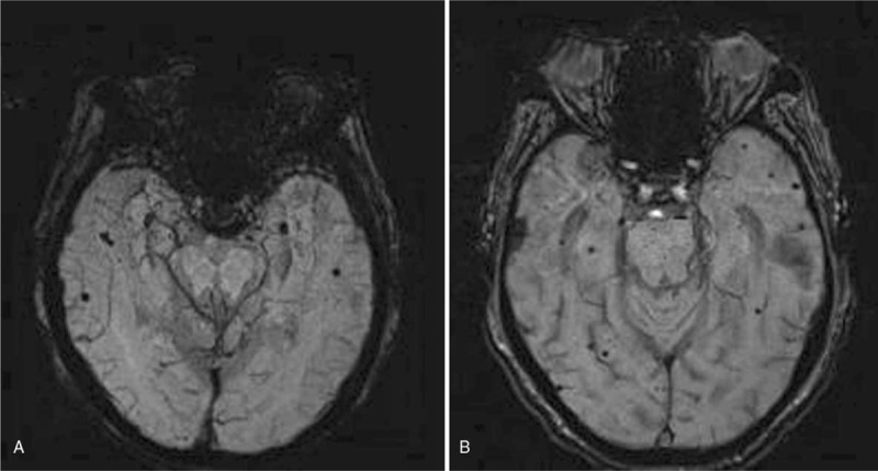
Forty patients with aMCI and AD respectively were recruited in the study, and 40 healthy subjects were taken as controls. Data were recorded using 3T MR scanner. We assessed the cerebral blood flow (CBF) in 11 different regions of interest, and counted number of microhemorrhages (MB) in 3 regions of brain lobes, bilateral basal ganglia/thalamus, and brain stem/cerebellum, and then investigated correlations between Montreal Cognitive Assessment (MoCA) scores, CBF, and susceptibility-weighted imaging (SWI) features in these 3 groups.
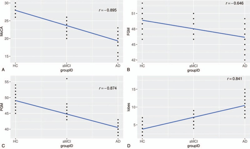
The results revealed that for AD patients, the MoCA scores and CBF values in frontal gray matter (FGM), occipital gray matter (OGM), temporal gray matter (TGM), parietal gray matter (PGM), hippocampus, anterior cingulate cortex (ACC), precuneus, posterior cingulate cortex (PCC), precuneus, basal ganglia and thalamus decreased compared with aMCI patients and control group, and significant difference was revealed among the 3 groups. While in cerebellum, statistical significance was only found between AD patients and control group. On SWI, the average numbers of hemorrhage in regions of lobes for AD patients were significantly higher than aMCI patients and control group. The same results occurred in the bilateral basal ganglia/thalamus. We further found the MoCA score was positively correlated with CBF, but negatively correlated with hypointense signal on SWI.
3D-pCASL and SWI have promising potential to be biomarkers for conversion from aMCI to AD in this process.
本研究旨在评估三维伪连续动脉自旋标记(3D-pcASL)和磁敏感加权成像(SWI)在此过程中作为遗忘型轻度认知障碍(aMCI)转化为阿尔茨海默病(AD)早期疾病敏感标志物的价值。
本研究招募了40例aMCI患者和40例AD患者,并选取40名健康受试者作为对照。使用3T磁共振成像扫描仪记录数据。我们评估了11个不同感兴趣区域的脑血流量(CBF),并计算了脑叶、双侧基底节/丘脑和脑干/小脑3个区域的微出血(MB)数量,然后研究了这3组中蒙特利尔认知评估(MoCA)评分、CBF和磁敏感加权成像(SWI)特征之间的相关性。
结果显示,与aMCI患者和对照组相比,AD患者的MoCA评分以及额叶灰质(FGM)、枕叶灰质(OGM)、颞叶灰质(TGM)、顶叶灰质(PGM)、海马体、前扣带回皮质(ACC)、楔前叶、后扣带回皮质(PCC)、楔前叶、基底节和丘脑的CBF值降低,3组之间存在显著差异。而在小脑方面,仅在AD患者和对照组之间发现统计学意义。在SWI上,AD患者脑叶区域的平均出血数明显高于aMCI患者和对照组。双侧基底节/丘脑也出现了相同的结果。我们进一步发现MoCA评分与CBF呈正相关,但与SWI上的低信号呈负相关。
3D-pCASL和SWI在此过程中作为aMCI转化为AD的生物标志物具有广阔的潜力。