Computer Science, State University of New York at Binghamton, Binghamton, NY, USA.
Harvard Medical School/Boston Children's Hospital, Boston, MA, USA.
J Alzheimers Dis. 2021;82(1):293-305. doi: 10.3233/JAD-210199.
This is the first longitudinal study to assess regional cerebral blood flow (rCBF) changes during the progression from normal control (NC) through mild cognitive impairment (MCI) and Alzheimer's disease (AD).
We aim to determine if perfusion MRI biomarkers, derived from our prior cross-sectional study, can predict the onset and cognitive decline of AD.
Perfusion MRIs using arterial spin labeling (ASL) were acquired in 15 stable-NC, 14 NC-to-MCI, 16 stable-MCI, and 18 MCI/AD-to-AD participants from the Cardiovascular Health Study (CHS) cognition study. Group comparisons, predictions of AD conversion and time to conversion, and Modified Mini-Mental State Examination (3MSE) from rCBF were performed.
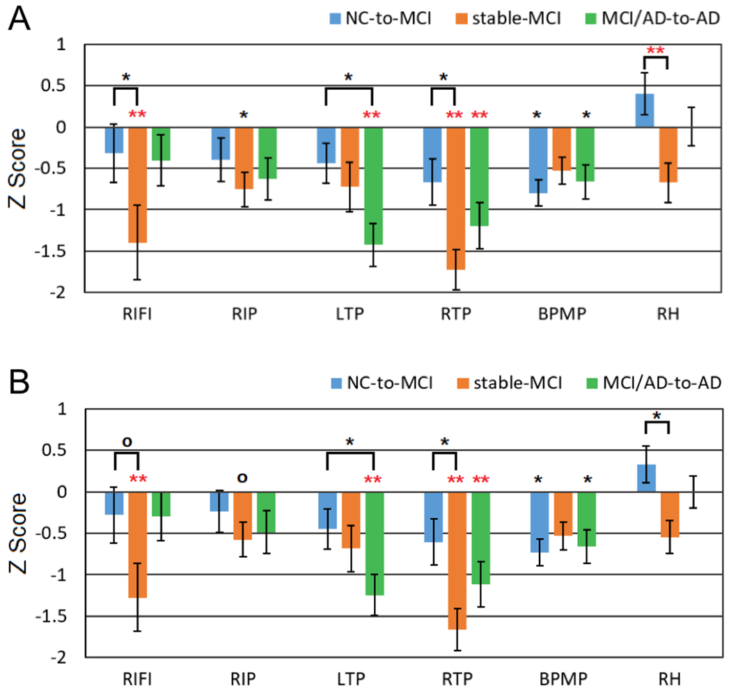
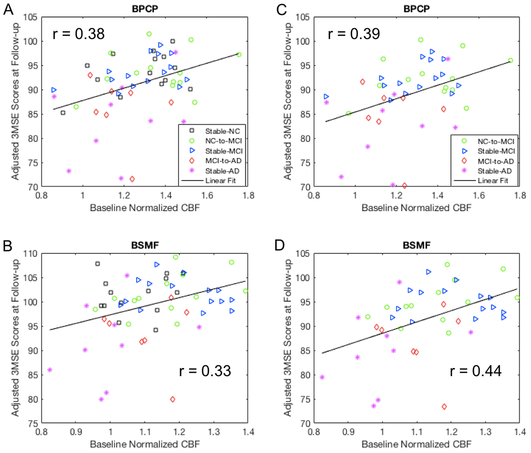
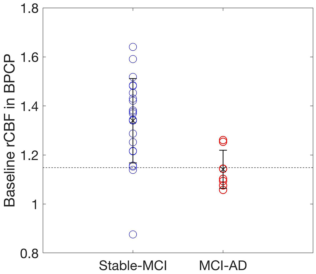
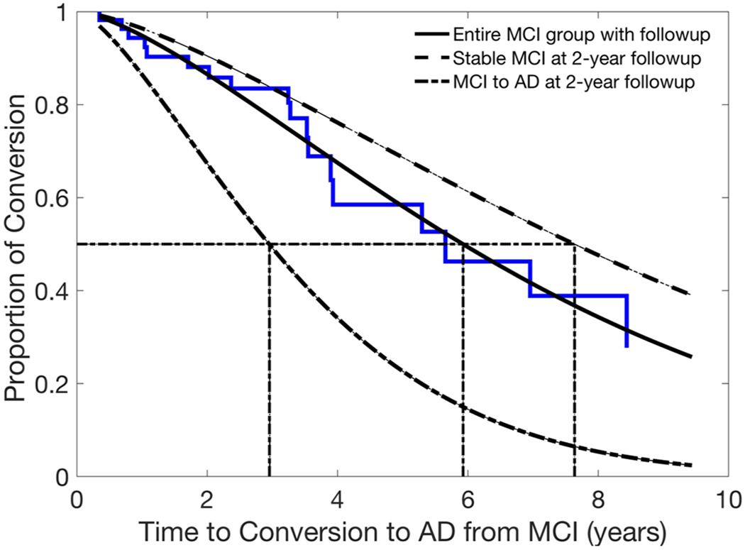
Compared to the stable-NC group: 1) the stable-MCI group exhibited rCBF decreases in the right temporoparietal (p = 0.00010) and right inferior frontal and insula (p = 0.0094) regions; and 2) the MCI/AD-to-AD group exhibited rCBF decreases in the bilateral temporoparietal regions (p = 0.00062 and 0.0035). Compared to the NC-to-MCI group, the stable-MCI group exhibited a rCBF decrease in the right hippocampus region (p = 0.0053). The baseline rCBF values in the posterior cingulate cortex (PCC) (p = 0.0043), bilateral superior medial frontal regions (BSMF) (p = 0.012), and left inferior frontal (p = 0.010) regions predicted the 3MSE scores for all the participants at follow-up. The baseline rCBF in the PCC and BSMF regions predicted the conversion and time to conversion from MCI to AD (p < 0.05; not significant after multiple corrections).
We demonstrated the feasibility of ASL in detecting rCBF changes in the typical AD-affected regions and the predictive value of baseline rCBF on AD conversion and cognitive decline.
这是第一项评估从正常对照(NC)经轻度认知障碍(MCI)到阿尔茨海默病(AD)进展过程中局部脑血流(rCBF)变化的纵向研究。
我们旨在确定先前的横断面研究中得出的灌注 MRI 生物标志物是否可预测 AD 的发病和认知下降。
采用动脉自旋标记(ASL)对来自心血管健康研究(CHS)认知研究的 15 例稳定 NC、14 例 NC 至 MCI、16 例稳定 MCI 和 18 例 MCI/AD 至 AD 患者进行灌注 MRI 检查。进行了组间比较、AD 转化率和达到转化率的时间以及 rCBF 的改良简易精神状态检查(3MSE)的预测。
与稳定 NC 组相比:1)稳定 MCI 组右侧颞顶叶(p = 0.00010)和右侧额下回和岛叶(p = 0.0094)区域 rCBF 降低;2)MCI/AD 至 AD 组双侧颞顶叶区域 rCBF 降低(p = 0.00062 和 0.0035)。与 NC 至 MCI 组相比,稳定 MCI 组右侧海马区域 rCBF 降低(p = 0.0053)。基线后扣带回皮质(PCC)(p = 0.0043)、双侧额上内侧回(BSMF)(p = 0.012)和左侧额下回(p = 0.010)的 rCBF 值预测了所有参与者随访时的 3MSE 评分。PCC 和 BSMF 区域的基线 rCBF 预测了从 MCI 到 AD 的转化和达到转化的时间(p < 0.05;经多次校正后无统计学意义)。
我们证明了 ASL 检测 AD 相关典型区域 rCBF 变化的可行性,以及基线 rCBF 对 AD 转化和认知下降的预测价值。