Laboratory of Surgical Oncology, Department of Surgery A, Tel-Aviv Sourasky Medical Center and The Sackler Faculty of Medicine, Tel-Aviv University, Tel-Aviv, Israel.
Casali Center for Applied Chemistry, Institute of Chemistry, The Hebrew University of Jerusalem, Jerusalem, Israel.
Sci Rep. 2019 Jun 12;9(1):8566. doi: 10.1038/s41598-019-45038-w.
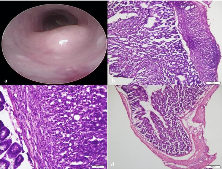
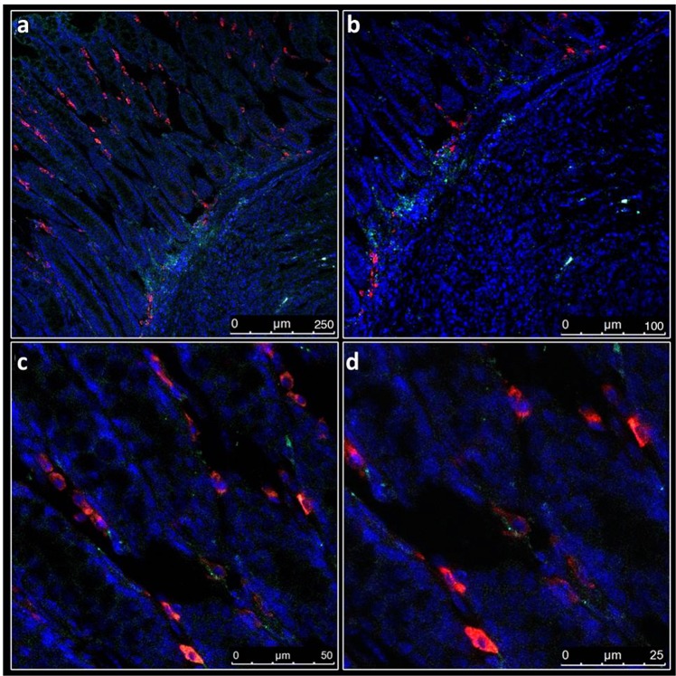
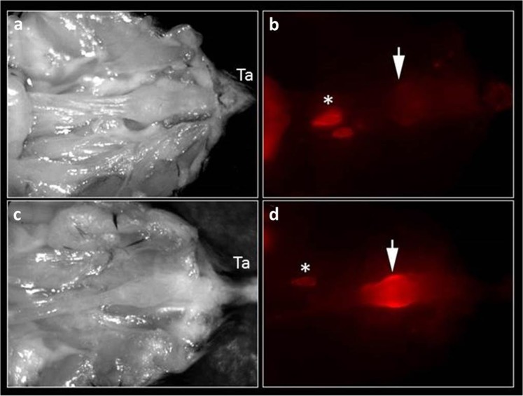
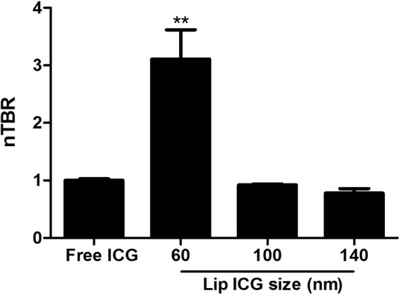
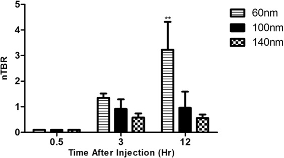
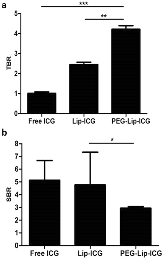
Localization of rectal tumors is a challenge in minimally invasive surgery due to the lack of tactile sensation. We had developed liposomal indocyanine green (Lip-ICG) for localization of rectal tumor. In this study we evaluated the effects of liposome size and lipid PEGylation on imaging. We used an endoscopically-guided orthotopic experimental rectal cancer model in which tumor fluorescence was determined at different time points after intravenous (i.v.) administration of Lip-ICG and PEGylated liposomes (PEG-Lip-ICG). Signal intensity was measured by tumor-to-background ratio (TBR), or normalized TBR (compared to TBR of free ICG). Fluorescence microscopy of tumor tissue was performed to determine fluorescence localization within the tissue and blood vessels. Liposomes of 60 nm showed an increased TBR compared with free ICG at 12 hours after i.v. injection: normalized TBR (nTBR) = 3.11 vs. 1, respectively (p = 0.006). Larger liposomes (100 nm and 140 nm) had comparable signal to free ICG (nTBR = 0.98 ± 0.02 and 0.78 ± 0.08, respectively), even when additional time points were examined (0.5, 3 and 24 hours). PEG-Lip- ICG were more efficient than Lip-ICG (TBR = 4.2 ± 0.18 vs. 2.5 ± 0.12, p < 0.01) presumably because of reduced uptake by the reticulo-endothelial system. ICG was found outside the capillaries in tumor margins. We conclude that size and lipid modification impact imaging intensity.
直肠肿瘤的定位在微创手术中是一个挑战,因为缺乏触觉。我们已经开发了用于直肠肿瘤定位的脂质体吲哚菁绿(Lip-ICG)。在这项研究中,我们评估了脂质体大小和脂质聚乙二醇化对成像的影响。我们使用了一种内窥镜引导的直肠原位实验性直肠癌模型,在该模型中,在静脉(i.v.)给予 Lip-ICG 和聚乙二醇化脂质体(PEG-Lip-ICG)后不同时间点测定肿瘤荧光。通过肿瘤与背景的比值(TBR)或与游离 ICG 的 TBR 进行归一化(normalized TBR)来测量信号强度。对肿瘤组织进行荧光显微镜检查,以确定组织和血管内的荧光定位。与游离 ICG 相比,60nm 的脂质体在静脉注射后 12 小时显示出更高的 TBR:归一化 TBR(nTBR)分别为 3.11 和 1(p=0.006)。较大的脂质体(100nm 和 140nm)与游离 ICG 的信号相似(nTBR 分别为 0.98±0.02 和 0.78±0.08),即使检查了更多的时间点(0.5、3 和 24 小时)。PEG-Lip-ICG 比 Lip-ICG 更有效(TBR=4.2±0.18 与 2.5±0.12,p<0.01),可能是因为网状内皮系统的摄取减少。ICG 被发现在肿瘤边缘的毛细血管外。我们的结论是,大小和脂质修饰会影响成像强度。