Witteveen M Emma, Flores Isadora Luana, Karssemakers Luc He, Bloemena Elisabeth
Department of Pathology, Amsterdam UMC, Vrije Universiteit Amsterdam, Amsterdam, The Netherlands.
Departamento de Odontologia Conservadora, Universidade Federal do Rio Grande do Sul, Porto Alegre, Brazil.
SAGE Open Med Case Rep. 2019 May 19;7:2050313X19849828. doi: 10.1177/2050313X19849828. eCollection 2019.
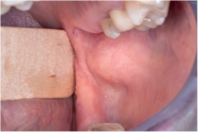
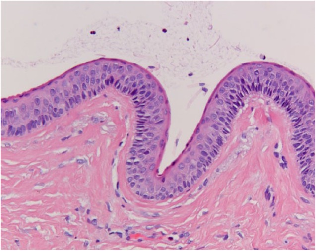
Odontogenic keratocysts make up 4%-12% of all odontogenic cysts. Most cysts are sporadic but sometimes they arise in the context of basal cell nevus syndrome (Gorlin syndrome). Most odontogenic keratocysts arise in the posterior region of the mandible, but they can occur anywhere in the jaw. In rare instances, they are located peripherally in the gingiva. Even more rare, they are found in the soft tissues of the mouth. There have been a few case reports and small case series of such peripheral odontogenic keratocysts. Some controversy exists as to whether these truly represent a peripheral counterpart of the intraosseous odontogenic keratocysts and if their origin is at all odontogenic. We hereby present two cases of peripheral odontogenic keratocysts, both being located in the soft tissue of the buccal mucosa, and review the literature on peripheral odontogenic keratocysts.
牙源性角化囊肿占所有牙源性囊肿的4%-12%。大多数囊肿为散发性,但有时会在基底细胞痣综合征(戈林综合征)的背景下出现。大多数牙源性角化囊肿发生在下颌骨后部区域,但也可发生于颌骨的任何部位。在罕见情况下,它们位于牙龈外周。更为罕见的是,它们见于口腔软组织。已有一些关于此类外周性牙源性角化囊肿的病例报告和小病例系列。对于这些是否真的代表骨内牙源性角化囊肿的外周对应物以及它们的起源是否完全为牙源性存在一些争议。我们在此报告两例外周性牙源性角化囊肿,均位于颊黏膜软组织,并复习外周性牙源性角化囊肿的相关文献。