Department of Radiology, West China Hospital, Sichuan University, 37# Guo Xue Xiang, Chengdu, Sichuan, 610041, China.
Department of Pathology, West China Hospital, Sichuan University, 37# Guo Xue Xiang, Chengdu, Sichuan, 610041, China.
Sci Rep. 2019 Jun 17;9(1):8657. doi: 10.1038/s41598-019-45144-9.
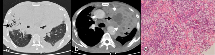
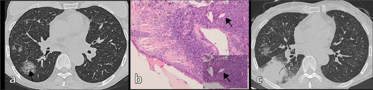
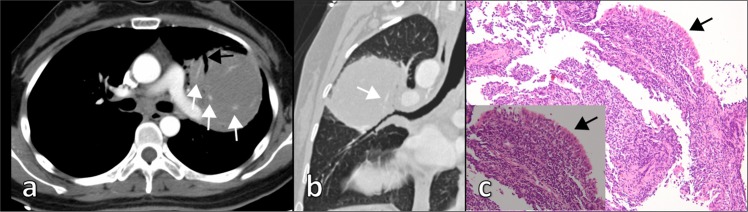
Pulmonary mucosa-associated lymphoid tissue (MALT) lymphoma is the most common primary pulmonary lymphoma. There are limited studies on imaging features of pulmonary MALT lymphoma. We present the computed tomography (CT) manifestations of pulmonary MALT lymphoma and the correlation between CT manifestations and clinical characteristics. Patients (n = 53) with histologically confirmed pulmonary MALT lymphoma who underwent chest CT scanning were retrospectively analyzed. Evaluated findings included distribution of pulmonary lesions, morphological pattern of appearance, contrast enhancement features, size, presence of thoracic lymphadenopathy, and secondary associated features. Pulmonary MALT lymphoma was observed in multiple (79%) and bilateral (66%) disease with random distribution (≥70%) of pulmonary lesions. The most frequent morphological pattern was consolidation (n = 33, 62%), followed by nodule (n = 23, 43%) and mass (n = 11, 21%). Common associated features were air bronchograms and bronchiectasis, especially cystic bronchiectasis and angiogram sign. Asymptomatic patients had less consolidation and bronchiectasis than did symptomatic patients. Cystic bronchiectasis was only observed in the symptomatic group. In conclusion, pulmonary MALT lymphoma manifests as diverse patterns on CT scans. Consolidation combined with cystic bronchiectasis was a characteristic late sign, which may assist in differential diagnosis. High-resolution CT images and multiplanar reconstruction techniques are helpful for accurately determining imaging manifestations.
肺黏膜相关淋巴组织(MALT)淋巴瘤是最常见的原发性肺淋巴瘤。关于肺 MALT 淋巴瘤的影像学特征研究有限。我们提出了肺 MALT 淋巴瘤的 CT 表现及其与临床特征的相关性。回顾性分析了经组织学证实为肺 MALT 淋巴瘤且接受胸部 CT 扫描的患者(n=53)。评估的发现包括肺部病变的分布、形态学表现、对比增强特征、大小、是否存在胸腔淋巴结病以及继发性相关特征。肺 MALT 淋巴瘤呈多发病灶(79%)和双侧病变(66%),且肺部病变呈随机分布(≥70%)。最常见的形态学表现为实变(n=33,62%),其次是结节(n=23,43%)和肿块(n=11,21%)。常见的相关特征是空气支气管征和支气管扩张,尤其是囊性支气管扩张和血管造影征。无症状患者的实变和支气管扩张较有症状患者少。囊性支气管扩张仅见于有症状组。总之,肺 MALT 淋巴瘤在 CT 扫描上表现为多种模式。伴有囊性支气管扩张的实变是一个特征性的晚期征象,有助于鉴别诊断。高分辨率 CT 图像和多平面重建技术有助于准确确定影像学表现。