Laboratoire de Dynamique des Interactions Membranaires Normales et Pathologiques, Université de Montpellier, CNRS-UMR5235, Montpellier, France.
LISM, Institut de Microbiologie de la Méditerranée, CNRS & Aix-Marseille Univ, Marseille, France.
PLoS Pathog. 2019 Jun 20;15(6):e1007812. doi: 10.1371/journal.ppat.1007812. eCollection 2019 Jun.
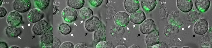
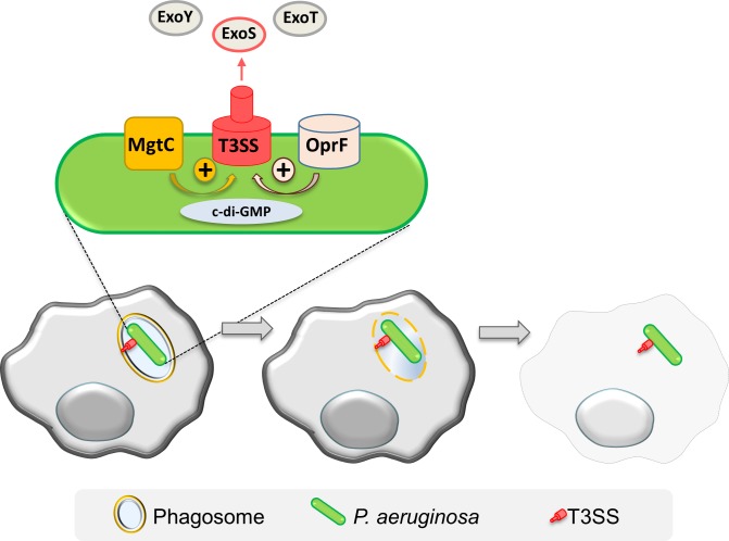
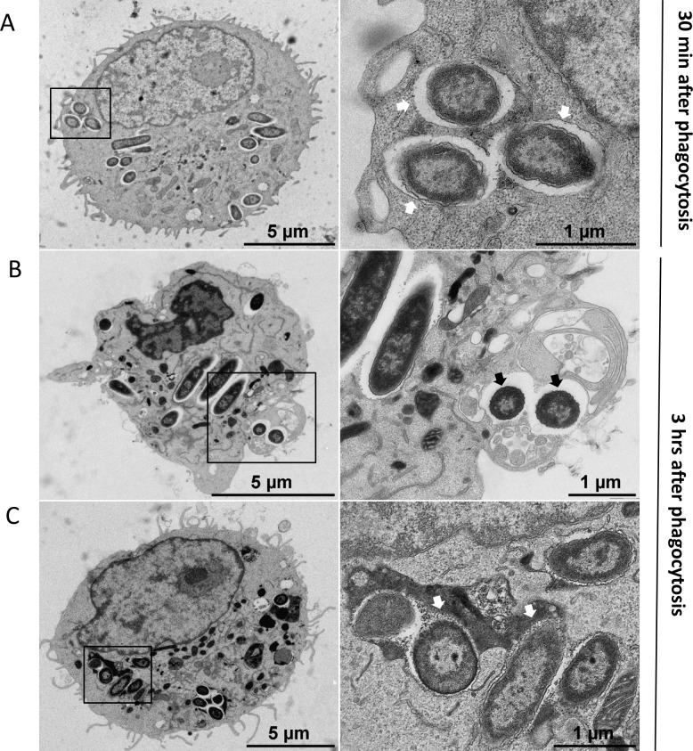
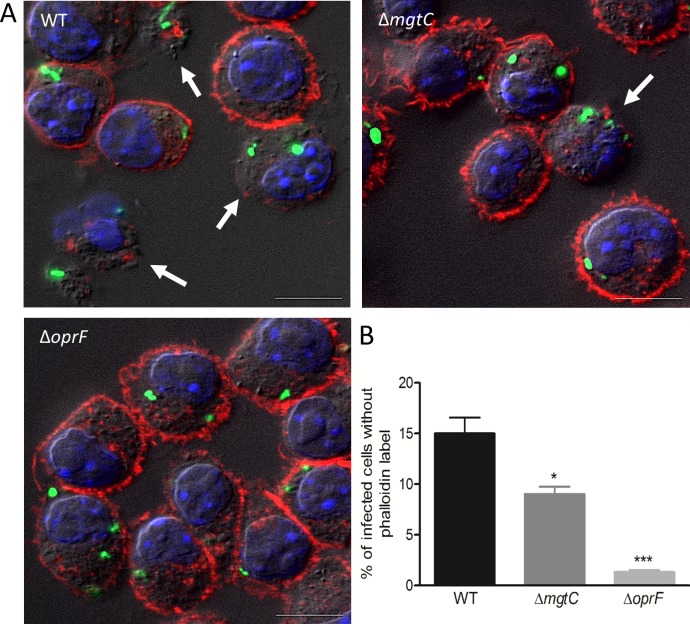
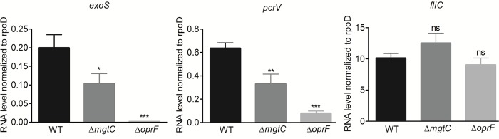
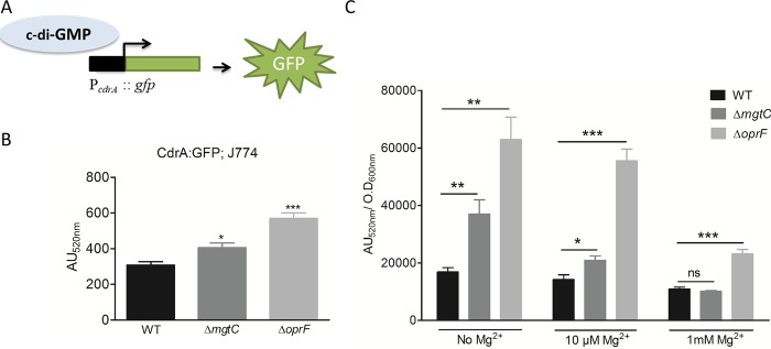
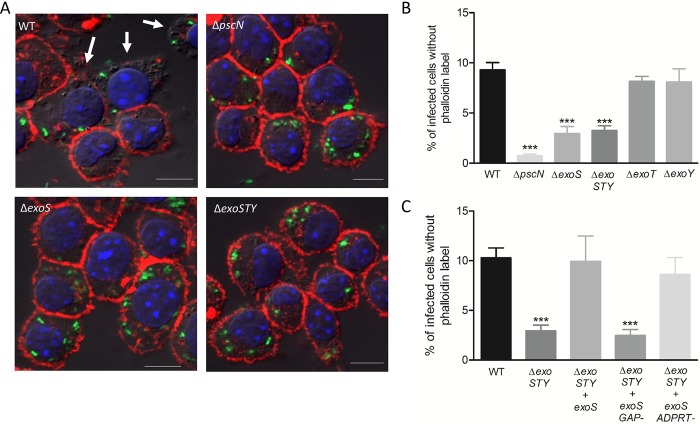
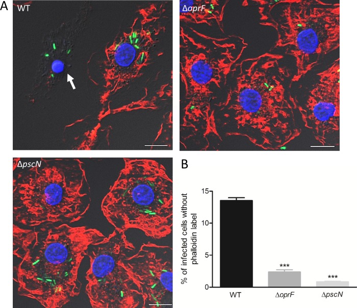
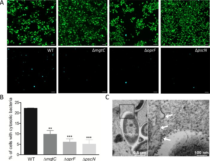
While considered solely an extracellular pathogen, increasing evidence indicates that Pseudomonas aeruginosa encounters intracellular environment in diverse mammalian cell types, including macrophages. In the present study, we have deciphered the intramacrophage fate of wild-type P. aeruginosa PAO1 strain by live and electron microscopy. P. aeruginosa first resided in phagosomal vacuoles and subsequently could be detected in the cytoplasm, indicating phagosomal escape of the pathogen, a finding also supported by vacuolar rupture assay. The intracellular bacteria could eventually induce cell lysis, both in a macrophage cell line and primary human macrophages. Two bacterial factors, MgtC and OprF, recently identified to be important for survival of P. aeruginosa in macrophages, were found to be involved in bacterial escape from the phagosome as well as in cell lysis caused by intracellular bacteria. Strikingly, type III secretion system (T3SS) genes of P. aeruginosa were down-regulated within macrophages in both mgtC and oprF mutants. Concordantly, cyclic di-GMP (c-di-GMP) level was increased in both mutants, providing a clue for negative regulation of T3SS inside macrophages. Consistent with the phenotypes and gene expression pattern of mgtC and oprF mutants, a T3SS mutant (ΔpscN) exhibited defect in phagosomal escape and macrophage lysis driven by internalized bacteria. Importantly, these effects appeared to be largely dependent on the ExoS effector, in contrast with the known T3SS-dependent, but ExoS independent, cytotoxicity caused by extracellular P. aeruginosa towards macrophages. Moreover, this macrophage damage caused by intracellular P. aeruginosa was found to be dependent on GTPase Activating Protein (GAP) domain of ExoS. Hence, our work highlights T3SS and ExoS, whose expression is modulated by MgtC and OprF, as key players in the intramacrophage life of P. aeruginosa which allow internalized bacteria to lyse macrophages.
虽然被认为仅是一种细胞外病原体,但越来越多的证据表明,铜绿假单胞菌在包括巨噬细胞在内的多种哺乳动物细胞类型中遇到细胞内环境。在本研究中,我们通过活细胞和电子显微镜解析了野生型铜绿假单胞菌 PAO1 株在巨噬细胞内的命运。铜绿假单胞菌首先存在于吞噬体小泡中,随后可以在细胞质中检测到,表明病原体逃避了吞噬体,这一发现也得到了空泡破裂测定的支持。胞内细菌最终可以诱导细胞裂解,无论是在巨噬细胞系还是原代人巨噬细胞中。最近发现,两种细菌因子 MgtC 和 OprF 对铜绿假单胞菌在巨噬细胞中的存活很重要,它们参与了细菌从吞噬体中的逃逸以及由胞内细菌引起的细胞裂解。引人注目的是,铜绿假单胞菌的 III 型分泌系统 (T3SS) 基因在 mgtC 和 oprF 突变体中的巨噬细胞中下调。一致地,两种突变体中的环二鸟苷酸 (c-di-GMP) 水平增加,为 T3SS 在巨噬细胞内的负调控提供了线索。与 mgtC 和 oprF 突变体的表型和基因表达模式一致,T3SS 突变体 (ΔpscN) 表现出吞噬体逃逸缺陷和由内化细菌驱动的巨噬细胞裂解缺陷。重要的是,这些效应似乎在很大程度上依赖于 ExoS 效应子,与已知的依赖于 T3SS 但不依赖于 ExoS 的由细胞外铜绿假单胞菌引起的巨噬细胞毒性不同。此外,发现由胞内铜绿假单胞菌引起的这种巨噬细胞损伤依赖于 ExoS 的 GTPase 激活蛋白 (GAP) 结构域。因此,我们的工作强调了 T3SS 和 ExoS,它们的表达受 MgtC 和 OprF 调节,是铜绿假单胞菌在巨噬细胞内生活中的关键因素,使内化细菌能够裂解巨噬细胞。