Saijo Hideto, Fujihara Yuko, Kanno Yuki, Hoshi Kazuto, Hikita Atsuhiko, Chung Ung-Il, Takato Tsuyoshi
Department of Oral and Maxillofacial Surgery, Faculty of Medicine, The University of Tokyo, Hongo 7-3-1, Bunkyoku, Tokyo 113-0033, Japan.
Division of Tissue Engineering at the University of Tokyo Hospital, Hongo 7-3-1, Bunkyoku, Tokyo 113-0033, Japan.
Regen Ther. 2016 Sep 20;5:72-78. doi: 10.1016/j.reth.2016.08.004. eCollection 2016 Dec.
Autologous, allogeneic, and artificial bones are clinically applied as graft materials for bone reconstruction, with each having their own advantages and disadvantages. Although artificial bones with various shapes are currently available, a product with a morphology that may be freely modified by operators has not yet been developed. In the present study, we developed a full custom-made artificial bone, and applied it to form the maxillofacial region. We herein report treatment outcomes.
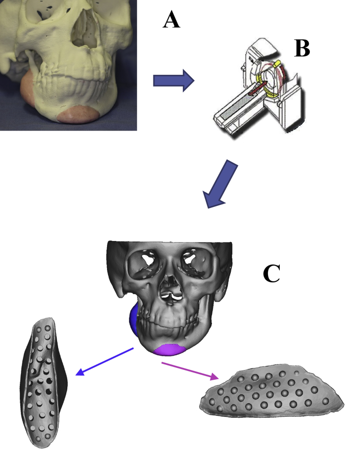
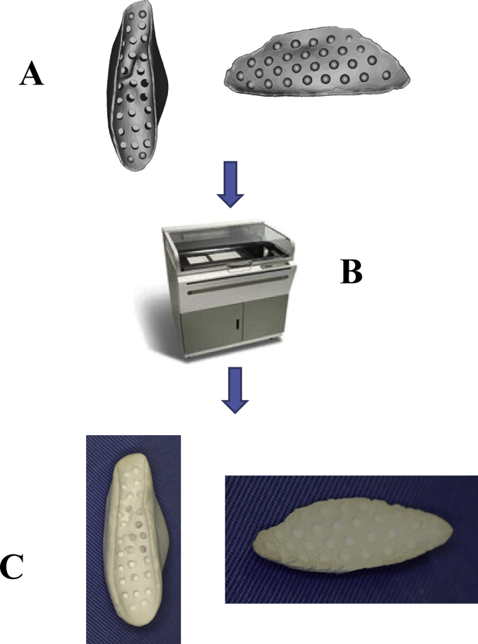
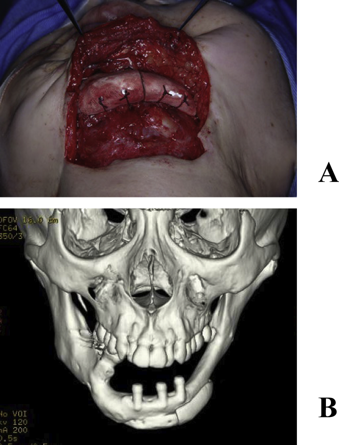
An artificial bone was prepared on a 3-dimensional solid model, and data of its shape was collected on CT. A full custom-made artificial bone was prepared by laminating α-tricalcium phosphate powder using an aqueous polysaccharide curing solution and the ink-jet powder-laminating device, Z406 3D Printer (DICO, USA). Subjects comprised patients who underwent maxillofacial plasty using this artificial bone between March 2006 and September 2009.
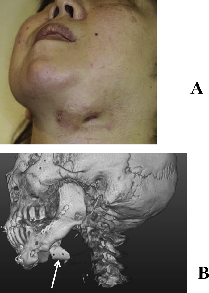
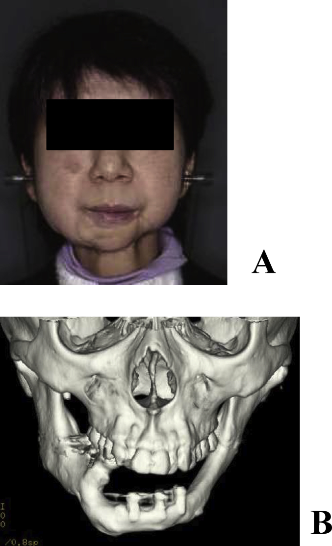
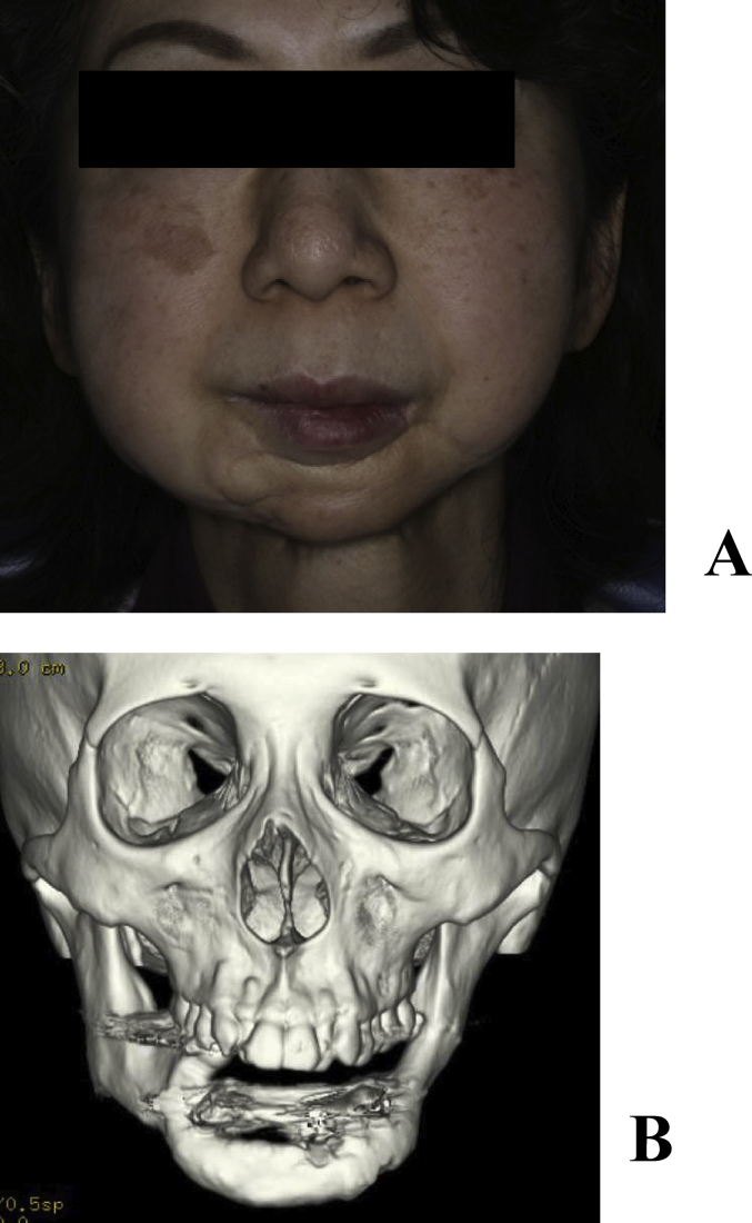
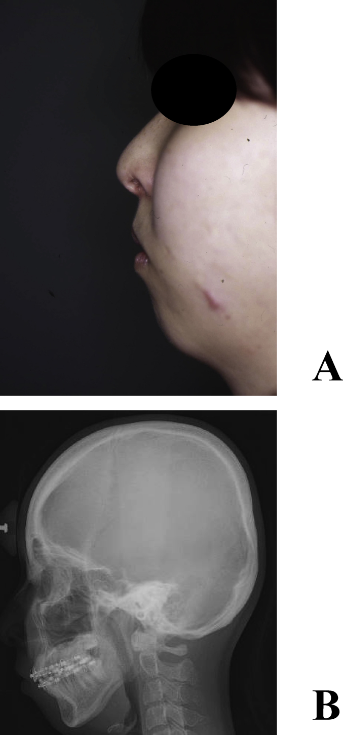
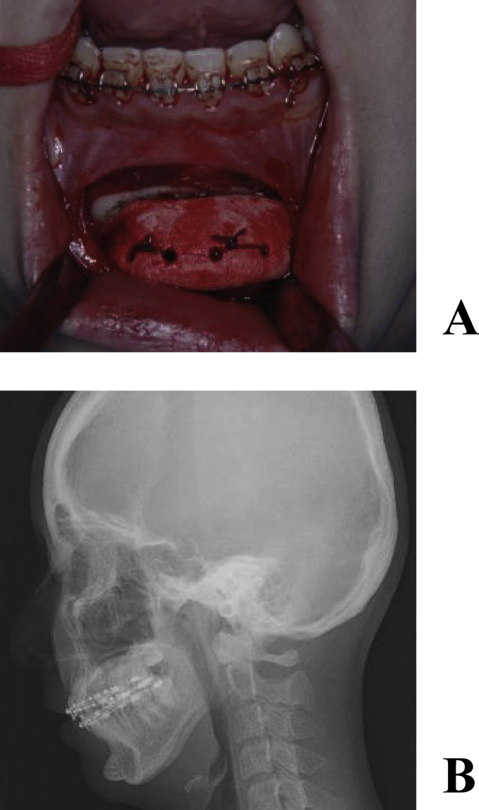
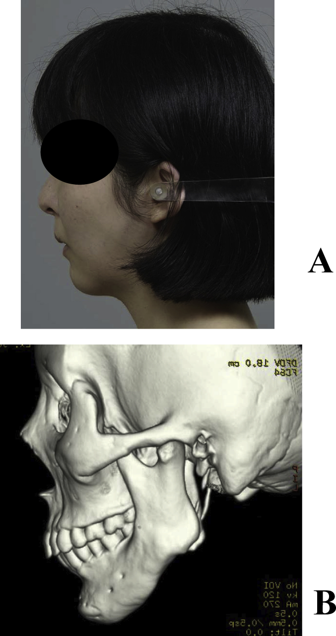
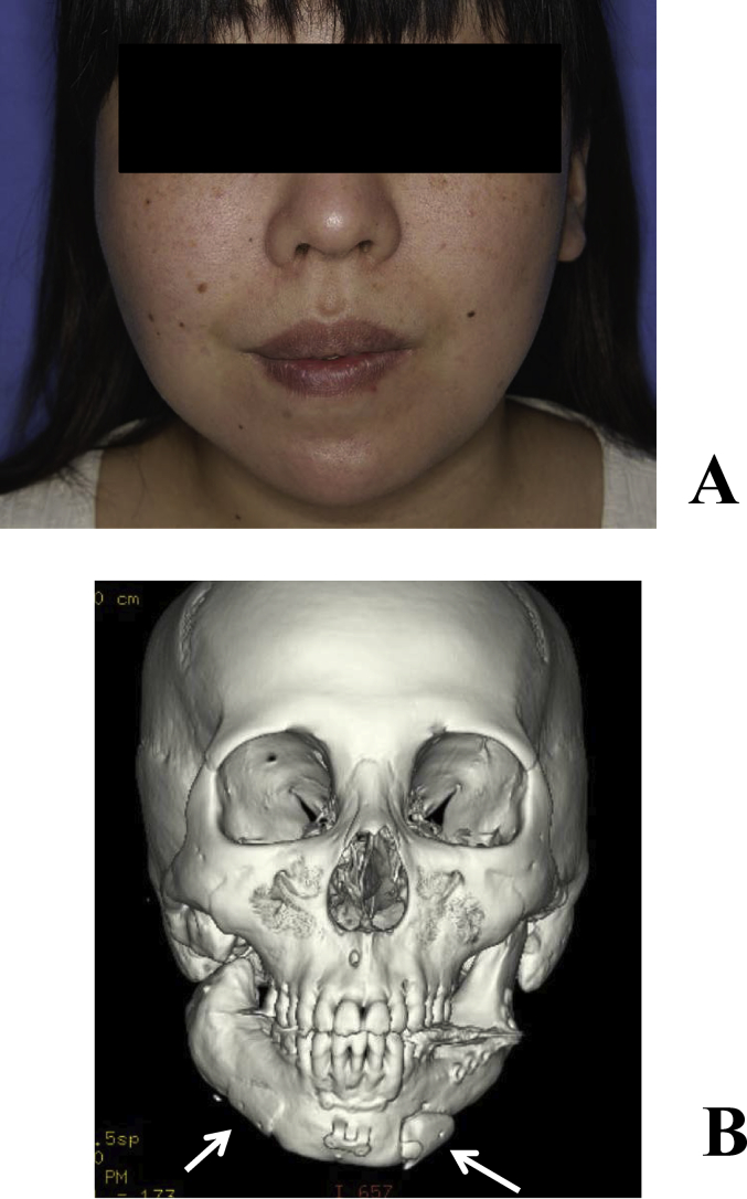
Maxillofacial plasty using the full custom-made artificial bone was applied to 23 regions in 20 patients (14 females and 6 males). The recipient region was the maxilla in 3, mandibular ramus in 13, mental region in 7, and frontal bone in 1. Postoperative courses were favorable in 18 out of the 23 regions; however, the fit was insufficient in 2 regions and the recipient regions were exposed within 1 year after surgery. Three regions were exposed 1 year or more after surgery.
We developed a novel reconstruction method using a full custom-made artificial bone. Its fit with the recipient bone was considered to be important, since an ill fit between the recipient and artificial bones potentially resulting in the artificial bone being detached. Therefore, fixation is important in order to prevent the detachment, and careful course observations are required when an ill fit is concerned during the follow-up period.
自体骨、异体骨和人工骨在临床上均被用作骨重建的移植材料,每种材料都有其自身的优缺点。尽管目前已有各种形状的人工骨,但尚未开发出一种其形态可由操作者自由修改的产品。在本研究中,我们开发了一种完全定制的人工骨,并将其应用于颌面区域的塑形。在此,我们报告治疗结果。
在三维实体模型上制备人工骨,并在CT上收集其形状数据。使用水性多糖固化溶液和喷墨粉末层压设备Z406 3D打印机(美国DICO公司)通过层压α-磷酸三钙粉末制备完全定制的人工骨。研究对象包括2006年3月至2009年9月间使用这种人工骨进行颌面整形的患者。
使用完全定制人工骨进行颌面整形应用于20例患者(14例女性和6例男性)的23个区域。受区为上颌骨3例、下颌支13例、颏部7例、额骨1例。23个区域中有18个术后病程良好;然而,2个区域贴合不充分,受区在术后1年内暴露。3个区域在术后1年或更长时间后暴露。
我们开发了一种使用完全定制人工骨的新型重建方法。其与受区骨的贴合被认为很重要,因为受区与人工骨之间贴合不佳可能导致人工骨脱落。因此,为防止脱落固定很重要,并且在随访期间当担心贴合不佳时需要仔细观察病程。