Department of Biomedical Engineering, The University of Alabama at Birmingham, Birmingham, Alabama, United States of America.
School of Medicine, The University of Alabama at Birmingham, Birmingham, Alabama, United States of America.
PLoS One. 2019 Jul 5;14(7):e0219442. doi: 10.1371/journal.pone.0219442. eCollection 2019.
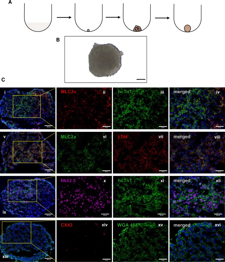
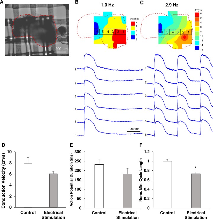
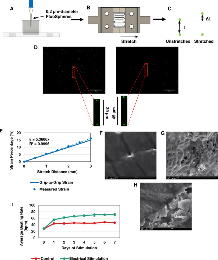
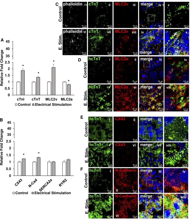
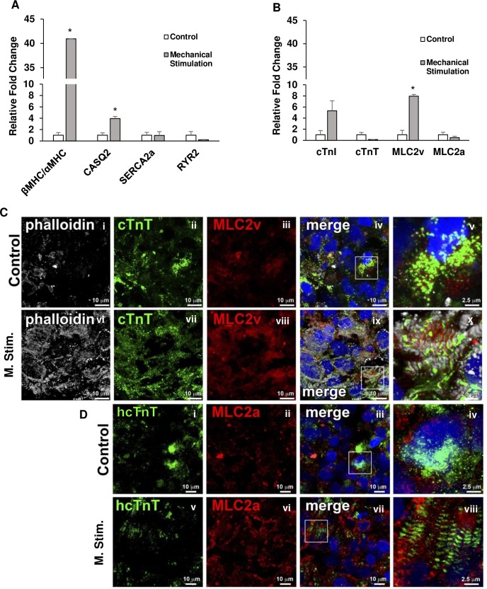
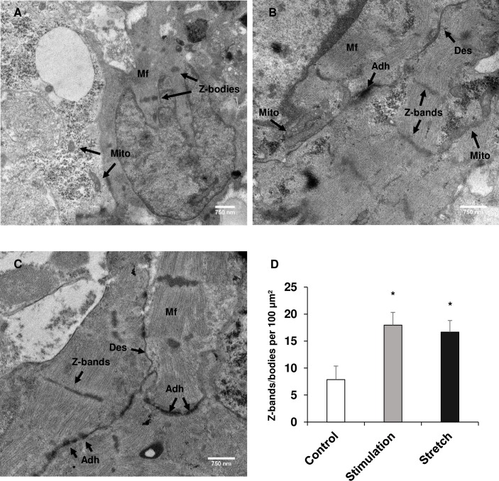
Functional myocardium derived from human induced pluripotent stem cells (hiPSCs) can be impactful for cardiac disease modeling, drug testing, and the repair of injured myocardium. However, when hiPSCs are differentiated into cardiomyocytes, they do not possess characteristics of mature myocytes which limits their application in these endeavors. We hypothesized that mechanical and electrical stimuli would enhance the maturation of hiPSC-derived cardiomyocyte (hiPSC-CM) spheroids on both a structural and functional level, potentially leading to a better model for drug testing as well as cell therapy. Spheroids were generated with hiPSC-CM. For inducing mechanical stimulation, they were placed in a custom-made device with PDMS channels and exposed to cyclic, uniaxial stretch. Spheroids were electrically stimulated in the C-Pace EP from IONOptix for 7 days. Following the stimulations, the spheroids were then analyzed for cardiomyocyte maturation. Both stimulated groups of spheroids possessed enhanced transcript and protein expressions for key maturation markers, such as cTnI, MLC2v, and MLC2a, along with improved ultrastructure of the hiPSC-CMs in both groups with enhanced Z-band/Z-body formation, fibril alignment, and fiber number. Optical mapping showed that spheroids exposed to electrical stimulation were able to capture signals at increasing rates of pacing up to 4 Hz, which failed in unstimulated spheroids. Our results clearly indicate that a significantly improved myocyte maturation can be achieved by culturing iPSC-CMs as spheroids and exposing them to cyclic, uniaxial stretch and electrical stimulation.
源自人诱导多能干细胞(hiPSCs)的功能性心肌可以对心脏病建模、药物测试和损伤心肌的修复产生重大影响。然而,当 hiPSCs 分化为心肌细胞时,它们不具有成熟心肌细胞的特征,这限制了它们在这些研究中的应用。我们假设机械和电刺激将在结构和功能水平上增强 hiPSC 衍生的心肌细胞(hiPSC-CM)球体的成熟,这可能会为药物测试以及细胞治疗提供更好的模型。使用 hiPSC-CM 生成球体。为了诱导机械刺激,将它们放置在具有 PDMS 通道的定制设备中,并暴露于周期性的单轴拉伸下。使用 IONOptix 的 C-Pace EP 对球体进行 7 天的电刺激。刺激后,然后分析球体以评估心肌细胞成熟度。两个刺激组的球体都表现出关键成熟标志物的转录和蛋白表达增强,例如 cTnI、MLC2v 和 MLC2a,以及两组 hiPSC-CMs 的超微结构得到改善,Z 带/Z 体形成、原纤维排列和纤维数量增加。光学映射显示,暴露于电刺激的球体能够以递增的起搏速率捕获信号,最高可达 4 Hz,而未受刺激的球体则无法做到这一点。我们的结果清楚地表明,通过培养 iPSC-CMs 作为球体并使其暴露于周期性的单轴拉伸和电刺激,可以实现显著改善的心肌细胞成熟。