Institute of Basic Medical Sciences, Chinese Academy of Medical Sciences & Peking Union Medical College, Beijing, 100005, China.
Materials Science and Devices Institute, Suzhou University of science and Technology, Suzhou, 215009, China.
Part Fibre Toxicol. 2019 Jul 12;16(1):30. doi: 10.1186/s12989-019-0314-4.
Iron oxide nanoparticles (IONPs) have been extensively studied in different biomedical fields. Recently, the non-cytotoxic concentration of IONPs induced cell-specific response raised concern of their safety. Endothelial cell exposure was unavoidable in their applications, while whether IONPs affect the phenotype of vascular endothelial cells is largely unknown. In this work, the effect of IONPs on endothelial-to-mesenchymal transition (EndMT) was investigated in vitro and in vivo.
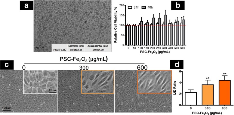
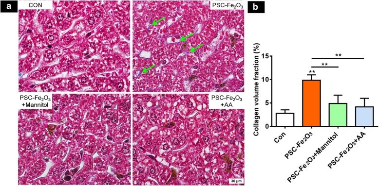
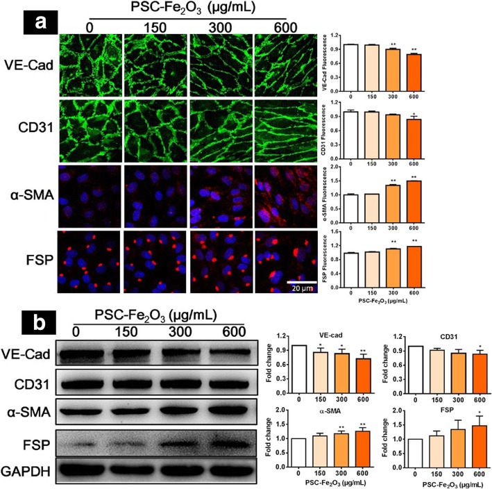
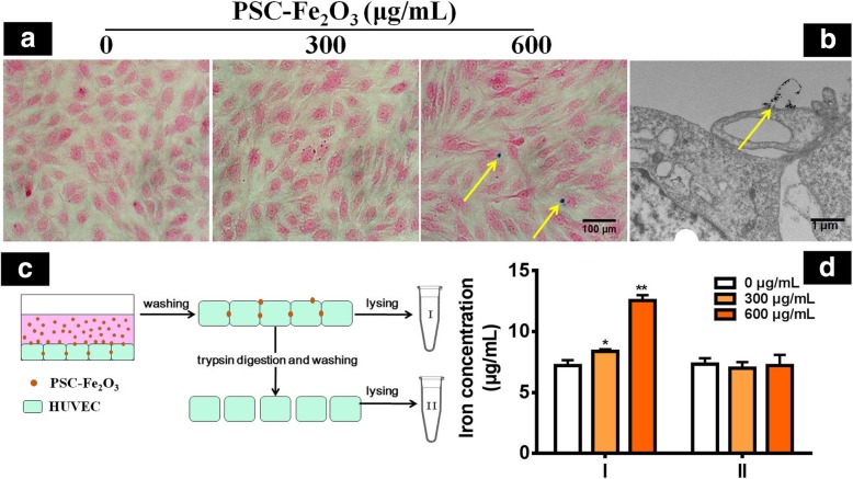
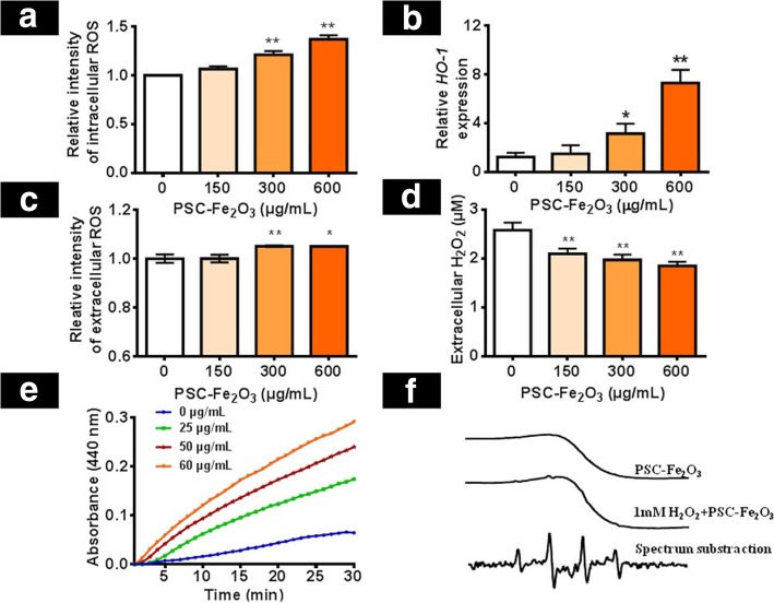
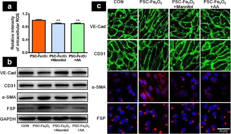
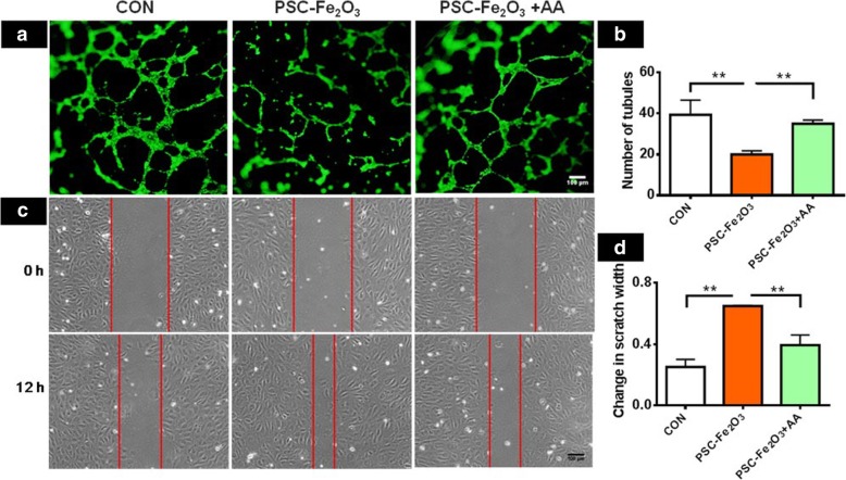
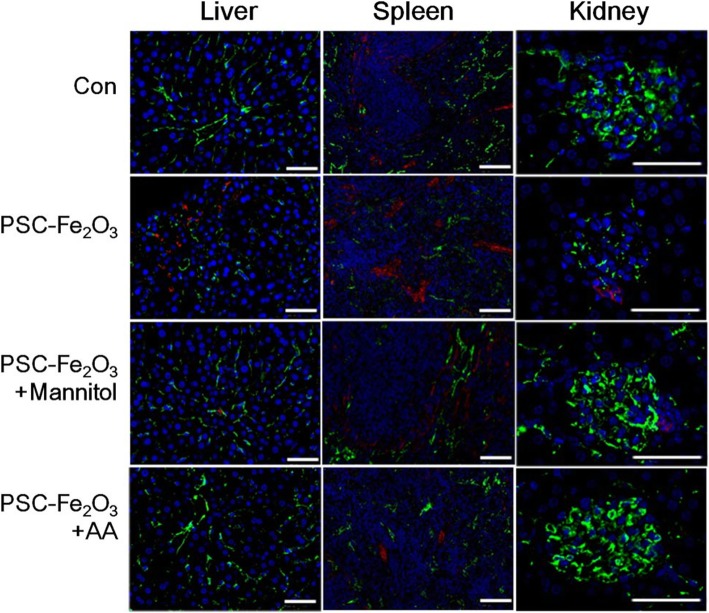
The incubation with γ-FeO nanoparticles modified with polyglucose sorbitol carboxymethyether (PSC-FeO) at non-cytotoxic concentration induced morphological changes of human umbilical vein endothelial cells (HUVECs) from cobblestone-like to spindle mesenchymal-like morphology, while PSC-FeO mostly stay in the culture medium and intercellular space. At the same time, the endothelial marker CD31 and VE-cadherin was decreased along with the inhibitory of angiogenesis properties of HUVEC. Meanwhile, the mesenchymal marker α-smooth muscle actin (α-SMA) and fibroblast specific protein (FSP) was up regulated significantly, and the migration ability of the cells was enhanced. When ROS scavenger mannitol or AA was supplemented, the EndMT was rescued. Results from the in vivo study showed that, expression of CD31 was decreased and α-SMA increased in the liver, spleen and kidney of mice given PSC-FeO, and the density of collagen fibers in the liver sinusoid of mice was increased. The supplementary mannitol or AA could reverse the degree of EndMT in the tissues. Mechanistic study in vitro indicated that the level of extracellular hydroxyl radicals (·OH) was up regulated significantly by PSC-FeO, which induced the response of intracellular ROS and resulted in the EndMT effect on HUVECs.
The PSC-FeO was capable of inducing EndMT in the endothelial cells at acutely non-cytotoxic dose due to its intrinsic peroxidase-like activity, though they were few taken up by endothelial cell. The EndMT effect on HUVEC can be rescued by ROS scavenger in vitro and in vivo.
氧化铁纳米粒子(IONPs)在不同的生物医学领域得到了广泛的研究。最近,研究发现 IONPs 在非细胞毒性浓度下诱导细胞特异性反应,这引起了人们对其安全性的关注。在其应用中,内皮细胞的暴露是不可避免的,而 IONPs 是否会影响血管内皮细胞的表型在很大程度上尚不清楚。在这项工作中,研究了 IONPs 在体外和体内对内皮细胞向间充质转化(EndMT)的影响。
在非细胞毒性浓度下孵育经过聚葡萄糖山梨醇羧甲基醚(PSC)修饰的γ-FeO 纳米粒子(PSC-FeO)会诱导人脐静脉内皮细胞(HUVEC)从鹅卵石样形态向梭形间充质样形态发生形态变化,而 PSC-FeO 主要停留在培养基和细胞间隙中。同时,内皮标志物 CD31 和 VE-钙粘蛋白的表达降低,伴随 HUVEC 血管生成能力受到抑制。同时,间充质标志物α-平滑肌肌动蛋白(α-SMA)和成纤维细胞特异性蛋白(FSP)显著上调,细胞迁移能力增强。当添加 ROS 清除剂甘露醇或 AA 时,EndMT 得到挽救。体内研究结果表明,给予 PSC-FeO 的小鼠肝脏、脾脏和肾脏中 CD31 的表达减少,α-SMA 增加,并且小鼠肝窦内皮细胞中的胶原纤维密度增加。补充甘露醇或 AA 可逆转组织中 EndMT 的程度。体外机制研究表明,PSC-FeO 显著上调细胞外羟基自由基(·OH)的水平,从而诱导细胞内 ROS 反应,导致 HUVEC 发生 EndMT 效应。
PSC-FeO 由于其内在的过氧化物酶样活性,在急性非细胞毒性剂量下能够诱导内皮细胞发生 EndMT,尽管它们很少被内皮细胞摄取。ROS 清除剂在体外和体内均可挽救 HUVEC 的 EndMT 效应。