• 文献检索
  • 文档翻译
  • 深度研究
  • 学术资讯
  • Suppr Zotero 插件Zotero 插件
  • 邀请有礼
  • 套餐&价格
  • 历史记录
应用&插件
Suppr Zotero 插件Zotero 插件浏览器插件Mac 客户端Windows 客户端微信小程序
定价
高级版会员购买积分包购买API积分包
服务
文献检索文档翻译深度研究API 文档MCP 服务
关于我们
关于 Suppr公司介绍联系我们用户协议隐私条款
关注我们

Suppr 超能文献

核心技术专利:CN118964589B侵权必究
粤ICP备2023148730 号-1Suppr @ 2026

文献检索

告别复杂PubMed语法,用中文像聊天一样搜索,搜遍4000万医学文献。AI智能推荐,让科研检索更轻松。

立即免费搜索

文件翻译

保留排版,准确专业,支持PDF/Word/PPT等文件格式,支持 12+语言互译。

免费翻译文档

深度研究

AI帮你快速写综述,25分钟生成高质量综述,智能提取关键信息,辅助科研写作。

立即免费体验

油酸可防止内皮细胞受到硅涂层超顺磁性氧化铁纳米颗粒(SPIONs)诱导的氧化应激和细胞死亡。

Oleic Acid Protects Endothelial Cells from Silica-Coated Superparamagnetic Iron Oxide Nanoparticles (SPIONs)-Induced Oxidative Stress and Cell Death.

机构信息

Biotechnical Faculty, University of Ljubljana, 1000 Ljubljana, Slovenia.

Department of Molecular and Biomedical Sciences, Jožef Stefan Institute, 1000 Ljubljana, Slovenia.

出版信息

Int J Mol Sci. 2022 Jun 23;23(13):6972. doi: 10.3390/ijms23136972.

DOI:10.3390/ijms23136972
PMID:35806014
原文链接:https://pmc.ncbi.nlm.nih.gov/articles/PMC9267005/
Abstract

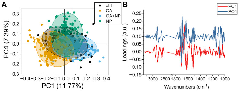
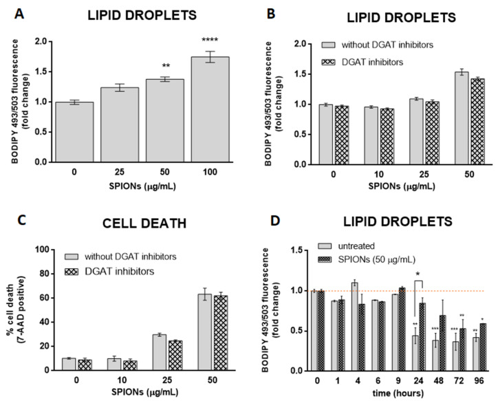
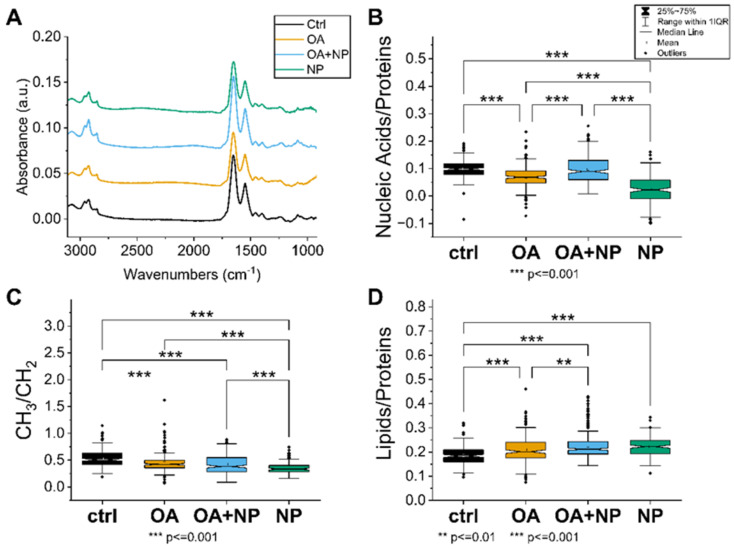
Superparamagnetic iron oxide nanoparticles (SPIONs) have great potential for use in medicine, but they may cause side effects due to oxidative stress. In our study, we investigated the effects of silica-coated SPIONs on endothelial cells and whether oleic acid (OA) can protect the cells from their harmful effects. We used viability assays, flow cytometry, infrared spectroscopy, fluorescence microscopy, and transmission electron microscopy. Our results show that silica-coated SPIONs are internalized by endothelial cells, where they increase the amount of reactive oxygen species (ROS) and cause cell death. Exposure to silica-coated SPIONs induced accumulation of lipid droplets (LD) that was not dependent on diacylglycerol acyltransferase (DGAT)-mediated LD biogenesis, suggesting that silica-coated SPIONs suppress LD degradation. Addition of exogenous OA promoted LD biogenesis and reduced SPION-dependent increases in oxidative stress and cell death. However, exogenous OA protected cells from SPION-induced cell damage even in the presence of DGAT inhibitors, implying that LDs are not required for the protective effect of exogenous OA. The molecular phenotype of the cells determined by Fourier transform infrared spectroscopy confirmed the destructive effect of silica-coated SPIONs and the ameliorative role of OA in the case of oxidative stress. Thus, exogenous OA protects endothelial cells from SPION-induced oxidative stress and cell death independent of its incorporation into triglycerides.

摘要

超顺磁性氧化铁纳米粒子(SPIONs)在医学中有很大的应用潜力,但由于氧化应激,它们可能会引起副作用。在我们的研究中,我们研究了硅涂层 SPIONs 对内皮细胞的影响,以及油酸(OA)是否可以保护细胞免受其有害影响。我们使用了细胞活力测定、流式细胞术、红外光谱、荧光显微镜和透射电子显微镜。我们的结果表明,硅涂层 SPIONs 被内皮细胞内化,在细胞内增加活性氧(ROS)的含量并导致细胞死亡。暴露于硅涂层 SPIONs 诱导脂滴(LD)的积累,这与二酰基甘油酰基转移酶(DGAT)介导的 LD 生物发生无关,表明硅涂层 SPIONs 抑制 LD 降解。添加外源性 OA 促进 LD 生物发生,并减少 SPION 依赖性氧化应激和细胞死亡的增加。然而,外源性 OA 即使在 DGAT 抑制剂存在的情况下也能保护细胞免受 SPION 诱导的细胞损伤,这表明 LD 对于外源性 OA 的保护作用不是必需的。傅里叶变换红外光谱法确定的细胞分子表型证实了硅涂层 SPIONs 的破坏作用和 OA 在氧化应激情况下的改善作用。因此,外源性 OA 可保护内皮细胞免受 SPION 诱导的氧化应激和细胞死亡,而与将其掺入三酸甘油脂无关。

https://cdn.ncbi.nlm.nih.gov/pmc/blobs/ab9f/9267005/228a33e6a110/ijms-23-06972-g006.jpg
https://cdn.ncbi.nlm.nih.gov/pmc/blobs/ab9f/9267005/10b97834e252/ijms-23-06972-g001.jpg
https://cdn.ncbi.nlm.nih.gov/pmc/blobs/ab9f/9267005/7fd759d5432f/ijms-23-06972-g002.jpg
https://cdn.ncbi.nlm.nih.gov/pmc/blobs/ab9f/9267005/661ba3b7b3da/ijms-23-06972-g003.jpg
https://cdn.ncbi.nlm.nih.gov/pmc/blobs/ab9f/9267005/f26b366b0859/ijms-23-06972-g004.jpg
https://cdn.ncbi.nlm.nih.gov/pmc/blobs/ab9f/9267005/b004d0b3d0b4/ijms-23-06972-g005.jpg
https://cdn.ncbi.nlm.nih.gov/pmc/blobs/ab9f/9267005/228a33e6a110/ijms-23-06972-g006.jpg

相似文献

1
Oleic Acid Protects Endothelial Cells from Silica-Coated Superparamagnetic Iron Oxide Nanoparticles (SPIONs)-Induced Oxidative Stress and Cell Death.油酸可防止内皮细胞受到硅涂层超顺磁性氧化铁纳米颗粒(SPIONs)诱导的氧化应激和细胞死亡。
Int J Mol Sci. 2022 Jun 23;23(13):6972. doi: 10.3390/ijms23136972.
2
Superparamagnetic iron-oxide nanoparticles mPEG350- and mPEG2000-coated: cell uptake and biocompatibility evaluation.超顺磁性氧化铁纳米颗粒 mPEG350- 和 mPEG2000 涂层:细胞摄取和生物相容性评价。
Nanomedicine. 2016 May;12(4):909-919. doi: 10.1016/j.nano.2015.12.371. Epub 2016 Jan 6.
3
Iodinated oil-loaded, fluorescent mesoporous silica-coated iron oxide nanoparticles for magnetic resonance imaging/computed tomography/fluorescence trimodal imaging.负载碘化油的荧光介孔二氧化硅包覆氧化铁纳米粒子用于磁共振成像/计算机断层扫描/荧光三模态成像。
Int J Nanomedicine. 2014 May 21;9:2527-38. doi: 10.2147/IJN.S59754. eCollection 2014.
4
Synthesis and Characterization of a Biocompatible Nanoplatform Based on Silica-Embedded SPIONs Functionalized with Polydopamine.基于二氧化硅包埋的聚多巴胺功能化超顺磁性氧化铁纳米颗粒的生物相容性纳米平台的合成与表征
ACS Biomater Sci Eng. 2023 Jan 9;9(1):303-317. doi: 10.1021/acsbiomaterials.2c00946. Epub 2022 Dec 9.
5
Innovative superparamagnetic iron-oxide nanoparticles coated with silica and conjugated with linoleic acid: Effect on tumor cell growth and viability.创新的二氧化硅包覆并与亚油酸共轭的超顺磁性氧化铁纳米颗粒:对肿瘤细胞生长和活力的影响。
Mater Sci Eng C Mater Biol Appl. 2017 Jul 1;76:439-447. doi: 10.1016/j.msec.2017.03.063. Epub 2017 Mar 10.
6
Increased endocytosis rate and enhanced lysosomal pathway of silica-coated superparamagnetic nanoparticles into M-HeLa cells compared with cultured primary motor neurons.与培养的原代运动神经元相比,硅涂层超顺磁纳米颗粒进入 M-HeLa 细胞的内吞速率增加,溶酶体途径增强。
Histochem Cell Biol. 2024 Jun;161(6):507-519. doi: 10.1007/s00418-024-02283-z. Epub 2024 Apr 10.
7
Biocompatible superparamagnetic core-shell nanoparticles for potential use in hyperthermia-enabled drug release and as an enhanced contrast agent.用于潜在的热疗增强药物释放和作为增强对比剂的生物相容性超顺磁核壳纳米粒子。
Nanotechnology. 2020 Sep 11;31(37):375102. doi: 10.1088/1361-6528/ab91f6. Epub 2020 May 11.
8
Heparin-Superparamagnetic Iron Oxide Nanoparticles for Theranostic Applications.肝素-超顺磁性氧化铁纳米粒子用于治疗诊断应用。
Molecules. 2022 Oct 21;27(20):7116. doi: 10.3390/molecules27207116.
9
Silica-coated superparamagnetic iron oxide nanoparticles targeting of EPCs in ischemic brain injury.靶向缺血性脑损伤中 EPC 的硅涂层超顺磁性氧化铁纳米颗粒。
Biomaterials. 2013 Jul;34(21):4982-92. doi: 10.1016/j.biomaterials.2013.03.030. Epub 2013 Apr 6.
10
Tubular electrosynthesis of silica-coated magnetite and evaluation of magnetic nanobiocatalyst efficacy during biomass hydrolysis.管状电合成二氧化硅包覆磁铁矿及磁性纳米生物催化剂在生物质水解过程中的催化效果评价。
Bioprocess Biosyst Eng. 2022 Aug;45(8):1311-1318. doi: 10.1007/s00449-022-02746-4. Epub 2022 Jul 25.

引用本文的文献

1
Comprehensive Analysis of the Potential Toxicity of Magnetic Iron Oxide Nanoparticles for Medical Applications: Cellular Mechanisms and Systemic Effects.磁性氧化铁纳米颗粒在医学应用中的潜在毒性的综合分析:细胞机制和系统效应。
Int J Mol Sci. 2024 Nov 8;25(22):12013. doi: 10.3390/ijms252212013.
2
Heating Capacity and Biocompatibility of Hybrid Nanoparticles for Magnetic Hyperthermia Treatment.用于磁热疗的混合纳米粒子的加热能力和生物相容性。
Int J Mol Sci. 2023 Dec 29;25(1):493. doi: 10.3390/ijms25010493.
3
Interaction of Nanomaterials with Cells and Tissues.

本文引用的文献

1
Ultrasmall iron oxide nanoparticles cause significant toxicity by specifically inducing acute oxidative stress to multiple organs.超小氧化铁纳米颗粒通过特异性诱导多个器官的急性氧化应激而引起显著毒性。
Part Fibre Toxicol. 2022 Mar 29;19(1):24. doi: 10.1186/s12989-022-00465-y.
2
A Versatile Interfacial Coassembly Method for Fabrication of Tunable Silica Shells with Radially Aligned Dual Mesopores on Diverse Magnetic Core Nanoparticles.一种用于在多种磁性核纳米颗粒上制备具有径向排列双介孔的可调谐二氧化硅壳的通用界面共组装方法。
ACS Appl Mater Interfaces. 2021 Jan 13;13(1):1883-1894. doi: 10.1021/acsami.0c17863. Epub 2021 Jan 4.
3
纳米材料与细胞和组织的相互作用。
Int J Mol Sci. 2023 Sep 4;24(17):13667. doi: 10.3390/ijms241713667.
4
Inflammatory, Oxidative Stress and Small Cellular Particle Response in HUVEC Induced by Debris from Endoprosthesis Processing.人工关节假体加工碎屑诱导人脐静脉内皮细胞的炎症、氧化应激及小细胞微粒反应
Materials (Basel). 2023 Apr 22;16(9):3287. doi: 10.3390/ma16093287.
5
Feeding Laying Ducks Leaves Increases the n-3 Fatty Acids Content and Decreases the n-6: n-3 PUFA Ratio in Egg Yolk without Affecting Laying Performance or Egg Quality.给产蛋鸭喂食树叶可增加蛋黄中n-3脂肪酸含量并降低n-6:n-3多不饱和脂肪酸比例,且不影响产蛋性能或蛋品质。
Foods. 2023 Jan 8;12(2):287. doi: 10.3390/foods12020287.
In vitro cytotoxicity study of superparamagnetic iron oxide and silica nanoparticles on pneumocyte organelles.
超顺磁性氧化铁和二氧化硅纳米粒子对肺细胞细胞器的体外细胞毒性研究。
Toxicol In Vitro. 2021 Apr;72:105071. doi: 10.1016/j.tiv.2020.105071. Epub 2020 Dec 29.
4
FTIR Spectroscopy to Reveal Lipid and Protein Changes Induced on Sperm by Capacitation: Bases for an Improvement of Sample Selection in ART.傅里叶变换红外光谱技术揭示顶体反应中精子脂质和蛋白质变化:ART 中样本选择改进的基础。
Int J Mol Sci. 2020 Nov 17;21(22):8659. doi: 10.3390/ijms21228659.
5
Lipid Droplets in Cancer.癌症中的脂滴
Rev Physiol Biochem Pharmacol. 2023;185:53-86. doi: 10.1007/112_2020_51.
6
High-sensitivity in vivo contrast for ultra-low field magnetic resonance imaging using superparamagnetic iron oxide nanoparticles.使用超顺磁性氧化铁纳米颗粒的超低场磁共振成像的高灵敏度体内造影
Sci Adv. 2020 Jul 17;6(29):eabb0998. doi: 10.1126/sciadv.abb0998. eCollection 2020 Jul.
7
Induction of lipid droplets in THP-1 macrophages by multi-walled carbon nanotubes in a diameter-dependent manner: A transcriptomic study.多壁碳纳米管以直径依赖方式诱导THP-1巨噬细胞中脂滴形成的转录组学研究
Toxicol Lett. 2020 Oct 10;332:65-73. doi: 10.1016/j.toxlet.2020.07.007. Epub 2020 Jul 7.
8
Differential response of liver sinusoidal endothelial cells and hepatocytes to oleic and palmitic acid revealed by Raman and CARS imaging.拉曼和 CARS 成像揭示油酸和棕榈酸对肝窦内皮细胞和肝细胞的差异反应。
Biochim Biophys Acta Mol Basis Dis. 2020 Jun 1;1866(6):165763. doi: 10.1016/j.bbadis.2020.165763. Epub 2020 Mar 10.
9
Toxicity assessment of superparamagnetic iron oxide nanoparticles in different tissues.不同组织中超顺磁氧化铁纳米颗粒的毒性评估。
Artif Cells Nanomed Biotechnol. 2020 Dec;48(1):443-451. doi: 10.1080/21691401.2019.1709855.
10
Oleic acid ameliorates palmitic acid induced hepatocellular lipotoxicity by inhibition of ER stress and pyroptosis.油酸通过抑制内质网应激和细胞焦亡改善棕榈酸诱导的肝细胞脂肪毒性。
Nutr Metab (Lond). 2020 Jan 30;17:11. doi: 10.1186/s12986-020-0434-8. eCollection 2020.