Laboratorio de Óptica, Instituto Universitario de Investigación en Óptica y Nanofísica, Universidad de Murcia, Campus de Espinardo (Ed. 34), 30100, Murcia, Spain.
Sci Rep. 2019 Jul 12;9(1):10121. doi: 10.1038/s41598-019-46568-z.
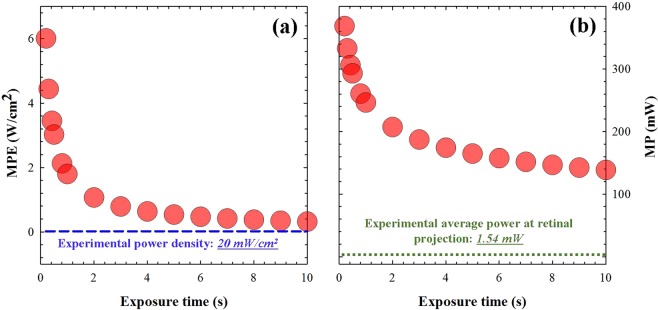
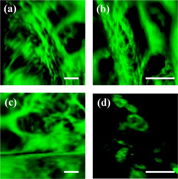
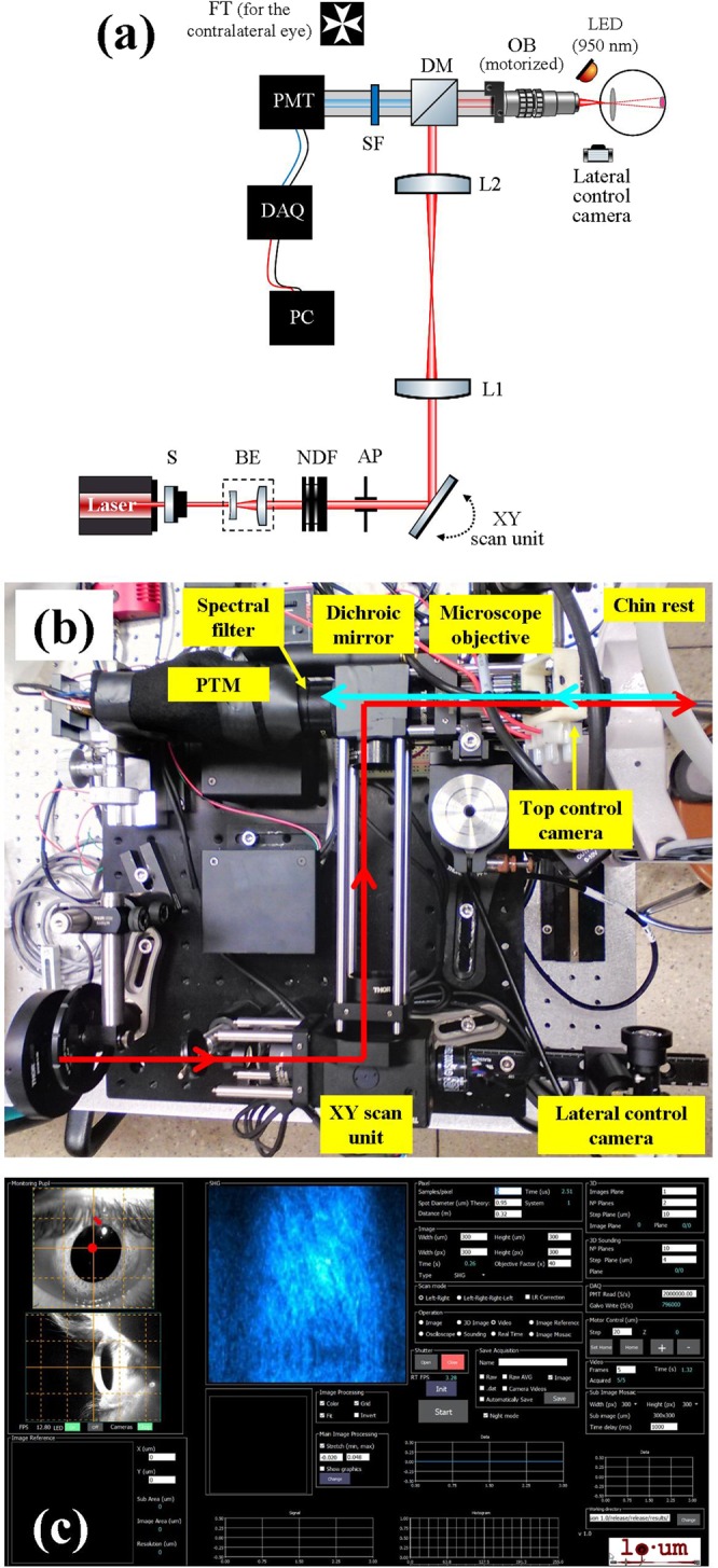
Two-photon (2P) microscopy is a powerful tool for imaging and exploring label-free biological tissues at high resolution. Although this type of microscopy has been demonstrated in ex vivo ocular tissues of both humans and animal models, imaging the human eye in vivo has always been challenging. This work presents a novel compact 2P microscope for non-contact imaging of the anterior part of the living human eye. The performance of the instrument was tested and the maximum permissible exposure to protect ocular tissues established. To the best of our knowledge, 2P images of the in vivo human cornea, the sclera and the trabecular meshwork are shown for the very first time. Acquired images are of enough quality to visualize collagen arrangement and morphological features of clinical interest. Future implementations of this technique may constitute a potential tool for early diagnosis of ocular diseases at submicron scale.
双光子(2P)显微镜是一种强大的工具,可用于高分辨率成像和探索无标记的生物组织。尽管这种类型的显微镜已经在人类和动物模型的离体眼部组织中得到了证明,但对活体人眼进行成像一直具有挑战性。本工作提出了一种新颖的紧凑型 2P 显微镜,用于对活体人眼的前节进行非接触式成像。测试了仪器的性能,并确定了保护眼部组织的最大允许暴露量。据我们所知,首次展示了活体人眼角膜、巩膜和小梁网的 2P 图像。所获得的图像质量足以可视化具有临床意义的胶原排列和形态特征。该技术的未来应用可能成为亚微米尺度早期诊断眼部疾病的潜在工具。