Department of Radiology, Molecular Imaging Program at Stanford, School of Medicine, Stanford University, Stanford, California.
Department of Radiation Oncology, Molecular Imaging Program at Stanford, School of Medicine, Stanford University, Stanford, California.
Cancer Res. 2019 Sep 15;79(18):4787-4797. doi: 10.1158/0008-5472.CAN-19-0530. Epub 2019 Jul 16.
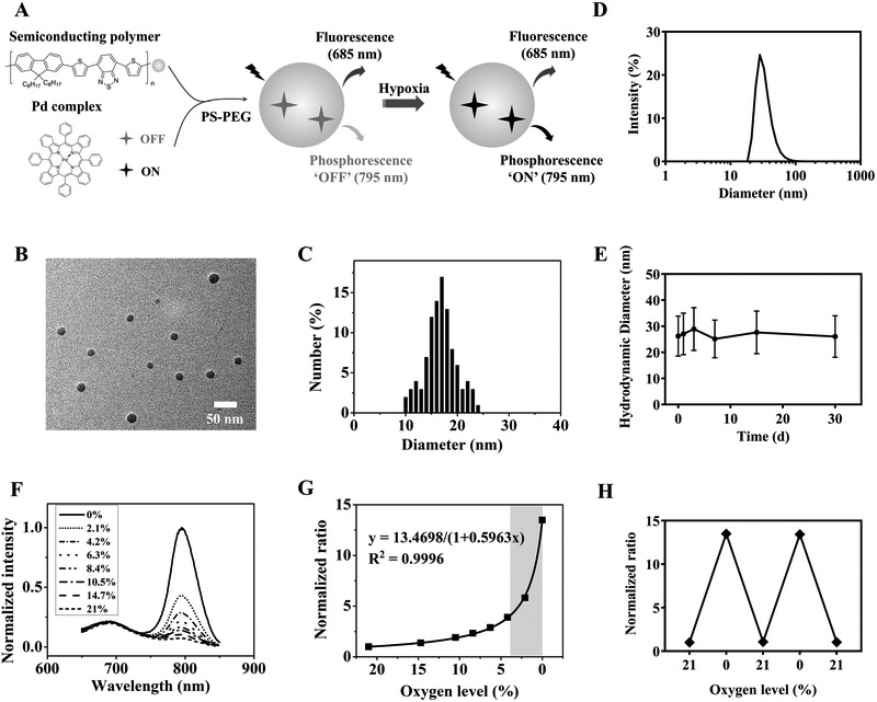
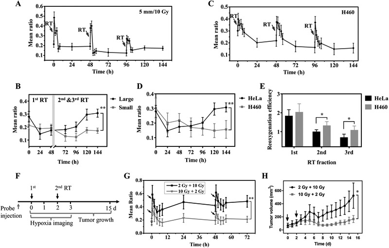
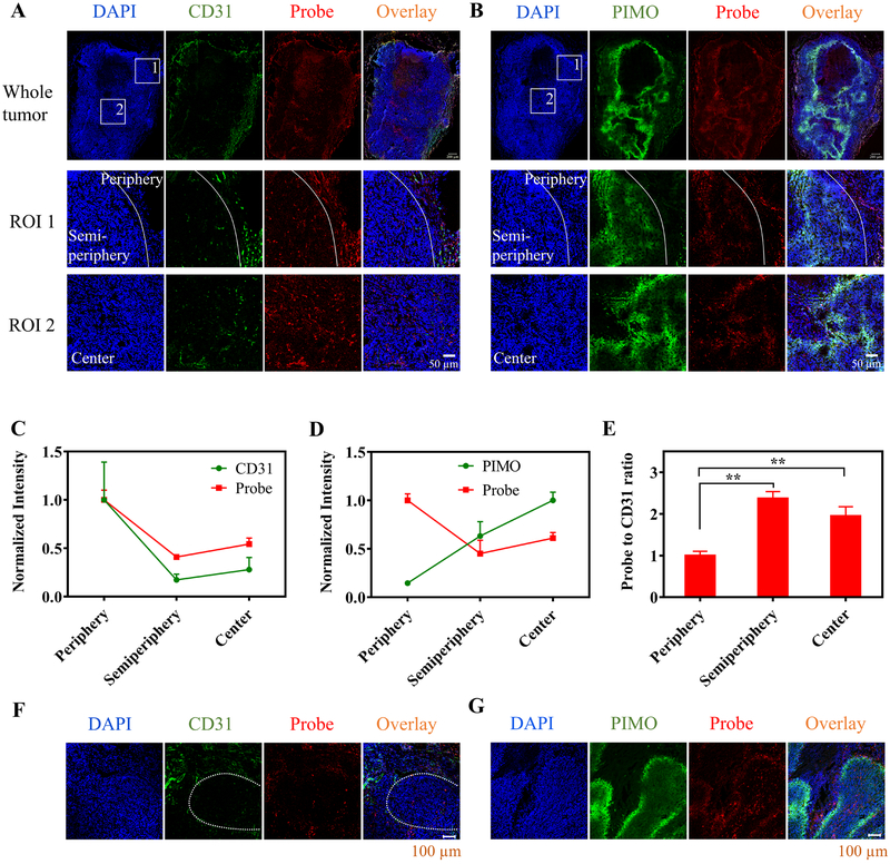
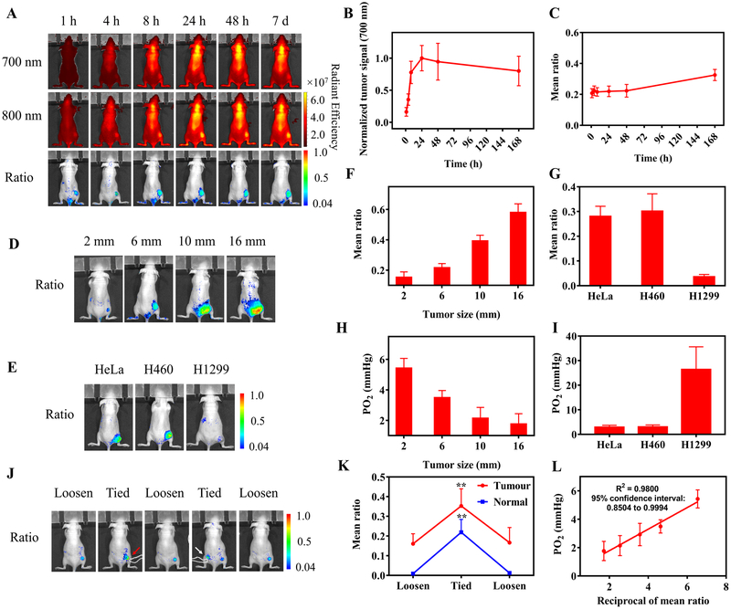
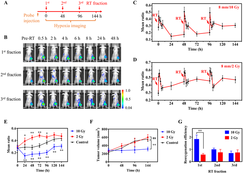
Hypoxia plays a key role in tumor resistance to radiotherapy. It is important to study hypoxia dynamics during radiotherapy to improve treatment planning and prognosis. Here, we describe a luminescent nanoprobe, composed of a fluorescent semiconducting polymer and palladium complex, for quantitative longitudinal imaging of tumor hypoxia dynamics during radiotherapy. The nanoprobe was designed to provide high sensitivity and reversible response for the subtle change in hypoxia over a narrow range (0-30 mmHg O), which spans the oxygen range where tumors have limited radiosensitivity. Following intravenous administration, the nanoprobe efficiently accumulated in and distributed across the tumor, including the hypoxic region. The ratio between emissions at 700 and 800 nm provided quantitative mapping of hypoxia across the entire tumor. The nanoprobe was used to image tumor hypoxia dynamics over 7 days during fractionated radiotherapy and revealed that high fractional dose (10 Gy) was more effective in improving tumor reoxygenation than low dose (2 Gy), and the effect tended to persist longer in smaller or more radiosensitive tumors. Our results also indicated the importance of the reoxygenation efficiency of the first fraction in the prediction of the radiation treatment outcome. In summary, this work has established a new nanoprobe for highly sensitive, quantitative, and longitudinal imaging of tumor hypoxia dynamics following radiotherapy, and demonstrated its value for assessing the efficacy of radiotherapy and radiation treatment planning. SIGNIFICANCE: This study presents a novel nanoagent for the visualization and quantification of tumor hypoxia.
缺氧在肿瘤对放射治疗的抵抗中起着关键作用。研究放射治疗过程中的缺氧动态变化对于改善治疗计划和预后非常重要。在这里,我们描述了一种由荧光半导体聚合物和钯配合物组成的发光纳米探针,用于定量纵向成像放射治疗过程中的肿瘤缺氧动力学。该纳米探针的设计提供了对缺氧微小变化的高灵敏度和可逆响应,其范围很窄(0-30 mmHg O),涵盖了肿瘤具有有限放射敏感性的氧气范围。静脉注射后,纳米探针能够有效地在肿瘤中积累并分布,包括缺氧区域。700nm 和 800nm 发射的比值提供了整个肿瘤缺氧的定量映射。该纳米探针用于在分割放射治疗期间对肿瘤缺氧动力学进行 7 天成像,结果表明高分次剂量(10 Gy)比低剂量(2 Gy)更有效地改善肿瘤再氧合,并且这种效果在较小或更敏感的肿瘤中持续时间更长。我们的结果还表明,第一分数的再氧合效率在预测放射治疗结果方面非常重要。总之,这项工作建立了一种新的纳米探针,用于高度敏感、定量和纵向成像放射治疗后的肿瘤缺氧动力学,并证明了其在评估放射治疗效果和放射治疗计划中的价值。意义:本研究提出了一种用于可视化和量化肿瘤缺氧的新型纳米剂。