• 文献检索
  • 文档翻译
  • 深度研究
  • 学术资讯
  • Suppr Zotero 插件Zotero 插件
  • 邀请有礼
  • 套餐&价格
  • 历史记录
应用&插件
Suppr Zotero 插件Zotero 插件浏览器插件Mac 客户端Windows 客户端微信小程序
定价
高级版会员购买积分包购买API积分包
服务
文献检索文档翻译深度研究API 文档MCP 服务
关于我们
关于 Suppr公司介绍联系我们用户协议隐私条款
关注我们

Suppr 超能文献

核心技术专利:CN118964589B侵权必究
粤ICP备2023148730 号-1Suppr @ 2026

文献检索

告别复杂PubMed语法,用中文像聊天一样搜索,搜遍4000万医学文献。AI智能推荐,让科研检索更轻松。

立即免费搜索

文件翻译

保留排版,准确专业,支持PDF/Word/PPT等文件格式,支持 12+语言互译。

免费翻译文档

深度研究

AI帮你快速写综述,25分钟生成高质量综述,智能提取关键信息,辅助科研写作。

立即免费体验

尸检时11.7特斯拉磁共振成像与偏振光成像显微镜用于测量人颈髓背根入髓区背根传入纤维的角度和方向

Post-mortem 11.7 Tesla Magnetic Resonance Imaging vs. Polarized Light Imaging Microscopy to Measure the Angle and Orientation of Dorsal Root Afferents in the Human Cervical Dorsal Root Entry Zone.

作者信息

Henssen Dylan Jozef Hendrik Augustinus, Weber Rosanna Christina, de Boef Jesse, Mollink Jeroen, Kozicz Tamas, Kurt Erkan, van Cappellen van Walsum Anne-Marie

机构信息

Department of Anatomy, Donders Institute for Brain, Cognition & Behavior, Radboud University Medical Center, Nijmegen, Netherlands.

Unit of Functional Neurosurgery, Department of Neurosurgery, Radboud University Medical Center, Nijmegen, Netherlands.

出版信息

Front Neuroanat. 2019 Jul 2;13:66. doi: 10.3389/fnana.2019.00066. eCollection 2019.

DOI:10.3389/fnana.2019.00066
PMID:31312124
原文链接:https://pmc.ncbi.nlm.nih.gov/articles/PMC6614433/
Abstract

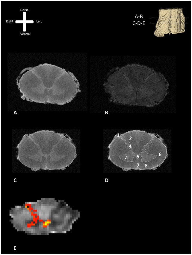
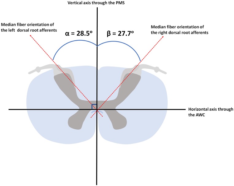
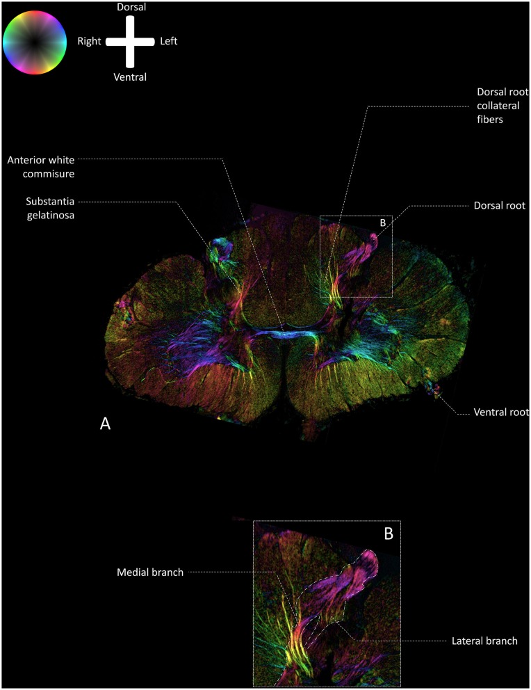
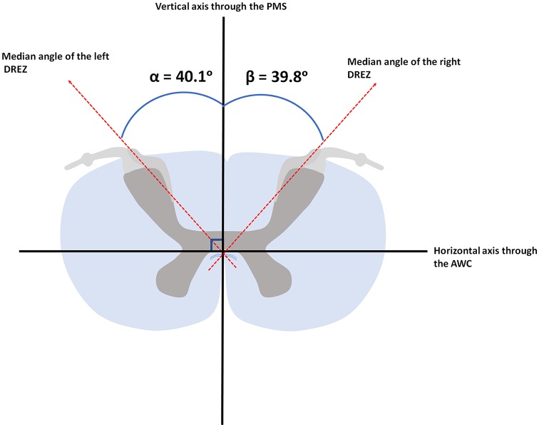
: Destruction of the afferents by dorsal root entry zone (DREZ) surgery may be an effective treatment of intractable neuropathic pain, though it remains a high-risk surgical intervention. Potential complications due to the lesioning of structures within the cervical spinal cord other than the DREZ can be minimized by accurate knowledge of the optimal insertion angle [i.e., the angle between the DREZ and the posterior median sulcus (PMS)]. The employed insertion angle was based on measurements between the DREZ and the PMS on post-mortem transverse slices. However, new, more sophisticated imaging techniques are currently available and are thought to yield higher spatial resolution and more accurate images. : This article measures the angle between the DREZ and the PMS on 11.7T post-mortem magnetic resonance images and compares these findings with polarized light imaging (PLI) microscopy images of the same specimens in order to quantify fiber orientation within the DREZ. : To visualize the anatomy of the cervical DREZ, magnetic resonance imaging (MRI), diffusion-weighted MRI (dMRI), probabilistic tractography, and PLI were performed on three post-mortem human cervical spinal cords at level C5-C6. The MR data was used to measure the angle between the DREZ and the PMS. MR images were complemented by probabilistic tractography results. Then, the orientation of fibers within the DREZ was quantified by use of PLI microscopy. : Median angle between the DREZ and the PMS, as measured on MR-images, was found to be 40.1° (ranging from 34.2° to 49.1°) and 39.8° (ranging from 31.1° to 47.8°) in the left and right hemicord, respectively. Median fiber orientation within the DREZ, as quantified by PLI, was 28.5° (ranging from 12.0° to 44.3°) and 27.7° (ranging from 8.5° to 38.1°) in the left and right hemicord, respectively. : Our study, which provides an improved understanding of the anatomy of the DREZ, the angle between the DREZ and the PMS and the median fiber orientation within the DREZ, could contribute to safer DREZ-lesioning surgery to treat chronic neuropathic pain in the future.

摘要

通过背根入髓区(DREZ)手术破坏传入神经可能是治疗顽固性神经性疼痛的一种有效方法,尽管它仍然是一种高风险的外科干预措施。通过准确了解最佳插入角度[即DREZ与后正中沟(PMS)之间的角度],可将因损伤颈髓内DREZ以外的结构而导致的潜在并发症降至最低。所采用的插入角度是基于对死后横切片上DREZ与PMS之间的测量。然而,目前已有更新的、更先进的成像技术,据认为这些技术能产生更高的空间分辨率和更精确的图像。本文在11.7T死后磁共振图像上测量DREZ与PMS之间的角度,并将这些结果与相同标本的偏振光成像(PLI)显微镜图像进行比较,以便量化DREZ内的纤维方向。为了可视化颈段DREZ的解剖结构,对三个死后人体颈髓C5 - C6节段进行了磁共振成像(MRI)、扩散加权MRI(dMRI)、概率性纤维束成像和PLI检查。利用MR数据测量DREZ与PMS之间的角度。MR图像辅以概率性纤维束成像结果。然后,通过PLI显微镜量化DREZ内纤维的方向。在MR图像上测量发现,左侧和右侧半脊髓中DREZ与PMS之间的中位角度分别为40.1°(范围为34.2°至49.1°)和39.8°(范围为31.1°至47.8°)。通过PLI量化,左侧和右侧半脊髓中DREZ内纤维的中位方向分别为28.5°(范围为12.0°至44.3°)和27.7°(范围为8.5°至38.1°)。我们的研究有助于更好地理解DREZ的解剖结构、DREZ与PMS之间的角度以及DREZ内纤维的中位方向,可能有助于未来更安全地进行DREZ毁损手术以治疗慢性神经性疼痛。

https://cdn.ncbi.nlm.nih.gov/pmc/blobs/d3de/6614433/a69d730930b5/fnana-13-00066-g0005.jpg
https://cdn.ncbi.nlm.nih.gov/pmc/blobs/d3de/6614433/34d2a91abeb7/fnana-13-00066-g0001.jpg
https://cdn.ncbi.nlm.nih.gov/pmc/blobs/d3de/6614433/4d006b2a69ab/fnana-13-00066-g0002.jpg
https://cdn.ncbi.nlm.nih.gov/pmc/blobs/d3de/6614433/576b209106fd/fnana-13-00066-g0003.jpg
https://cdn.ncbi.nlm.nih.gov/pmc/blobs/d3de/6614433/e68cf2eaef1d/fnana-13-00066-g0004.jpg
https://cdn.ncbi.nlm.nih.gov/pmc/blobs/d3de/6614433/a69d730930b5/fnana-13-00066-g0005.jpg

相似文献

1
Post-mortem 11.7 Tesla Magnetic Resonance Imaging vs. Polarized Light Imaging Microscopy to Measure the Angle and Orientation of Dorsal Root Afferents in the Human Cervical Dorsal Root Entry Zone.尸检时11.7特斯拉磁共振成像与偏振光成像显微镜用于测量人颈髓背根入髓区背根传入纤维的角度和方向
Front Neuroanat. 2019 Jul 2;13:66. doi: 10.3389/fnana.2019.00066. eCollection 2019.
2
Correlation of preoperative MRI with the long-term outcomes of dorsal root entry zone lesioning for brachial plexus avulsion pain.术前磁共振成像与臂丛神经撕脱伤疼痛的背根入髓区毁损术长期疗效的相关性
J Neurosurg. 2016 May;124(5):1470-8. doi: 10.3171/2015.2.JNS142572. Epub 2015 Sep 25.
3
Dorsal Root Entry Zone Lesioning for the Treatment of Pain After Brachial Plexus Avulsion Injury: 2-Dimensional Operative Video and Technical Report.经皮脊神经根入区切断术治疗臂丛神经撕脱伤后疼痛:二维手术视频及技术报告。
Oper Neurosurg (Hagerstown). 2022 Jun 1;22(6):e252-e258. doi: 10.1227/ons.0000000000000149. Epub 2022 Apr 14.
4
Microsurgical anatomy of dorsal root entry zone of brachial plexus.臂丛神经后根入髓区的显微外科解剖学
Microsurgery. 2008;28(1):17-20. doi: 10.1002/micr.20438.
5
Modified dorsal root entry zone lesioning for intractable pain relief in patients with root avulsion injury.改良背根入髓区毁损术用于神经根撕脱伤患者的顽固性疼痛缓解
J Neurosurg Spine. 2017 Aug;27(2):178-184. doi: 10.3171/2017.1.SPINE16234. Epub 2017 Jun 2.
6
Changes in spontaneous dorsal horn potentials after dorsal root entry zone lesioning in patients with pain after brachial plexus avulsion.臂丛神经撕脱伤后疼痛患者背根入髓区损伤后背角自发电位的变化
J Int Med Res. 2012;40(4):1499-506. doi: 10.1177/147323001204000429.
7
Retrospective Study of 57 Patients Submitted to Dorsal Root Entry Zone Lesioning by Radiofrequency for Brachial Plexus Avulsion Pain.对57例因臂丛神经撕脱伤疼痛接受射频背根入髓区毁损术患者的回顾性研究。
World Neurosurg. 2023 Sep;177:e466-e471. doi: 10.1016/j.wneu.2023.06.077. Epub 2023 Jun 24.
8
Dorsal Root Entry Zone Lesioning Following Unresponsive Spinal Cord Stimulation for Post-Traumatic Neuropathic Pain.脊髓刺激治疗创伤后神经病理性疼痛无效后行背根入区损毁术。
World Neurosurg. 2023 Oct;178:e300-e306. doi: 10.1016/j.wneu.2023.07.048. Epub 2023 Jul 18.
9
Spinal and Nucleus Caudalis Dorsal Root Entry Zone Lesioning for Chronic Pain: Efficacy and Outcomes.脊髓与尾状核背根入髓区损伤治疗慢性疼痛:疗效与结果
World Neurosurg. 2015 Aug;84(2):494-504. doi: 10.1016/j.wneu.2015.04.025. Epub 2015 Apr 18.
10
[Biomechanical characteristics of spinal cord tissue--basis for the development of modifications of the DREZ (dorsal root entry zone) operation].脊髓组织的生物力学特性——脊髓背根入髓区(DREZ)手术改良发展的基础
Acta Chir Iugosl. 2004;51(4):59-64. doi: 10.2298/aci0404059s.

引用本文的文献

1
Clinical Application of Diffusion Tensor Imaging for a Brachial Plexus Injury.扩散张量成像在臂丛神经损伤中的临床应用
Diagnostics (Basel). 2022 Jul 11;12(7):1687. doi: 10.3390/diagnostics12071687.

本文引用的文献

1
Ex vivo visualization of the trigeminal pathways in the human brainstem using 11.7T diffusion MRI combined with microscopy polarized light imaging.采用 11.7T 弥散 MRI 结合显微镜偏光成像技术对人脑干三叉神经通路进行体外可视化研究。
Brain Struct Funct. 2019 Jan;224(1):159-170. doi: 10.1007/s00429-018-1767-1. Epub 2018 Oct 6.
2
The Associated Factors and Clinical Features of Neuropathic Pain After Brachial Plexus Injuries: A Cross-sectional Study.臂丛神经损伤后神经病理性疼痛的相关因素及临床特征:一项横断面研究。
Clin J Pain. 2017 Nov;33(11):1030-1036. doi: 10.1097/AJP.0000000000000493.
3
Non-parametric representation and prediction of single- and multi-shell diffusion-weighted MRI data using Gaussian processes.
使用高斯过程对单壳和多壳扩散加权磁共振成像数据进行非参数表示和预测。
Neuroimage. 2015 Nov 15;122:166-76. doi: 10.1016/j.neuroimage.2015.07.067. Epub 2015 Jul 30.
4
Neuropathic pain after brachial plexus avulsion--central and peripheral mechanisms.臂丛神经撕脱伤后的神经性疼痛——中枢和外周机制
BMC Neurol. 2015 May 4;15:73. doi: 10.1186/s12883-015-0329-x.
5
Usefulness of screening tools in the evaluation of long-term effectiveness of DREZ lesioning in the treatment of neuropathic pain after brachial plexus injury.筛查工具在评估脊髓背根入髓区毁损术治疗臂丛神经损伤后神经性疼痛长期疗效中的作用
BMC Neurol. 2014 Dec 9;14:225. doi: 10.1186/s12883-014-0225-9.
6
Improving diffusion-weighted imaging of post-mortem human brains: SSFP at 7 T.改善死后人类大脑的扩散加权成像:7T下的稳态自由进动序列
Neuroimage. 2014 Nov 15;102 Pt 2:579-89. doi: 10.1016/j.neuroimage.2014.08.014. Epub 2014 Aug 13.
7
Layer-specific diffusion weighted imaging in human primary visual cortex in vitro.体外人初级视皮层的层特异性弥散加权成像。
Cortex. 2013 Oct;49(9):2569-82. doi: 10.1016/j.cortex.2012.11.015. Epub 2012 Dec 12.
8
High-resolution fiber tract reconstruction in the human brain by means of three-dimensional polarized light imaging.利用三维偏振光成像技术对人脑进行高分辨率纤维束重建。
Front Neuroinform. 2011 Dec 30;5:34. doi: 10.3389/fninf.2011.00034. eCollection 2011.
9
Microstructural analysis of human white matter architecture using polarized light imaging: views from neuroanatomy.利用偏光成像技术分析人类白质结构的微观结构:神经解剖学观点。
Front Neuroinform. 2011 Nov 11;5:28. doi: 10.3389/fninf.2011.00028. eCollection 2011.
10
FSL.束流输送系统。
Neuroimage. 2012 Aug 15;62(2):782-90. doi: 10.1016/j.neuroimage.2011.09.015. Epub 2011 Sep 16.