Bajaj Sahil, Raikes Adam C, Smith Ryan, Vanuk John R, Killgore William D S
Social, Cognitive and Affective Neuroscience Laboratory (SCAN Lab), Department of Psychiatry, College of Medicine, University of Arizona, Tucson, AZ, United States.
The Laureate Institute for Brain Research (LIBR), Tulsa, OK, United States.
Front Psychiatry. 2019 Jun 21;10:445. doi: 10.3389/fpsyt.2019.00445. eCollection 2019.
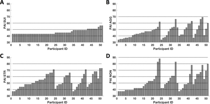
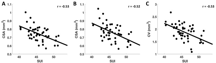
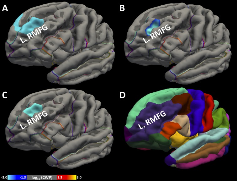
Suicidal ideation (SUI) can occur in the absence of concomitant psychiatric diagnoses, and even normal levels can be problematic among individuals experiencing excess stress or lack of social support. The objective of this study was to investigate the neuroanatomical basis of SUI in non-clinical human populations who are within the normal limits of SUI, after accounting for elevated stress and perceived lack of social support. Neuroanatomical data were collected from 55 healthy individuals (mean age 30.9 ± 8.1 years, 27 females) whose depression severity levels were below the criteria. Measures of SUI, aggression, stress, non-support, and treatment rejection were collected from the treatment-consideration scales (TCS) of the Personality Assessment Inventory (PAI). Correlations between standardized SUI scores and three brain morphometry measures, vertex wise cortical thickness (CT), cortical surface area (CSA), and cortical volume (CV), were estimated for each participant, controlling for age, sex, intracranial volume, and the remaining TCS measures. We observed a significant negative association between scores on SUI and both CSA and CV (cluster-forming threshold of < 0.005, clusterwise threshold of < 0.05, corrected for multiple comparisons) within the left rostral middle frontal gyrus. Our findings suggest that greater CSA and CV within the dorsolateral prefrontal cortex are associated with reduced SUI in a non-clinical population with mild levels of stress and perceived lack of social support. Because the dorsolateral prefrontal cortex has been broadly linked to cognitive reappraisal, self-critical thoughts, and emotional regulation, greater CSA and CV within these regions may lead to better mental health by protecting healthy individuals from engaging in SUI during periods of stress and perceived insufficient social support. As our data consisted of only healthy individuals with non-clinical levels of SUI, further investigation will be necessary to explore the neural basis of SUI in populations who may be at greater risk of future suicidal behavior.
自杀意念(SUI)可能在没有并发精神疾病诊断的情况下出现,甚至在经历过度压力或缺乏社会支持的个体中,即使处于正常水平也可能存在问题。本研究的目的是在考虑到压力升高和感知到的社会支持不足的情况下,调查处于自杀意念正常范围内的非临床人群中自杀意念的神经解剖学基础。从55名健康个体(平均年龄30.9±8.1岁,27名女性)收集神经解剖学数据;这些个体的抑郁严重程度低于标准。从人格评估量表(PAI)的治疗考虑量表(TCS)中收集自杀意念、攻击性、压力、缺乏支持和治疗拒绝的测量数据。针对每位参与者,在控制年龄、性别、颅内体积和其余TCS测量值的情况下,估计标准化自杀意念分数与三种脑形态测量指标(顶点皮质厚度(CT)、皮质表面积(CSA)和皮质体积(CV))之间的相关性。我们观察到,在左侧额中回喙部,自杀意念得分与CSA和CV均呈显著负相关(聚类形成阈值<0.005,聚类水平阈值<0.05,经多重比较校正)。我们的研究结果表明,在压力水平较轻且感知到缺乏社会支持的非临床人群中,背外侧前额叶皮质内更大的CSA和CV与自杀意念减少有关。由于背外侧前额叶皮质与认知重评、自我批评性思维和情绪调节广泛相关,这些区域内更大的CSA和CV可能通过在压力期和感知到社会支持不足时保护健康个体不产生自杀意念,从而带来更好的心理健康。由于我们的数据仅包括自杀意念处于非临床水平的健康个体,因此有必要进行进一步研究,以探索未来可能有更高自杀行为风险人群中自杀意念的神经基础。