Department of Mechanical and Industrial Engineering, College of Engineering; Biomedical Research Center (BRC), Qatar University, Doha, Qatar.
Biomedical Research Center (BRC), Qatar University; College of Medicine, Qatar University, Doha, Qatar.
Bosn J Basic Med Sci. 2020 Feb 5;20(1):140-148. doi: 10.17305/bjbms.2019.4372.
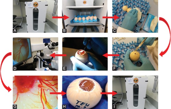
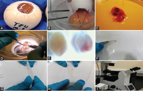
Cancer metastasis is the leading cause of cancer-related mortality worldwide. To date, several in vitro methodologies have been developed to understand the mechanisms of cancer metastasis and to screen various therapeutic agents against it. Nevertheless, mimicking an in vivo microenvironment in vitro is not possible; while in vivo experiments are complex, expensive and bound with several regulatory requirements. Herein, we report a novel in ovo model that relies on chicken embryo to investigate cancer cell invasion and metastasis to various organs of the body. In this model, we directly injected green fluorescent protein (GFP) expressing cancer cells to the heart of chicken embryo at 3 days of incubation, then monitored cell migration to various organs. To this end, we used a simple tissue processing technique to achieve rapid imaging and quantification of invasive cells. We were able to clearly observe the migration of GFP expressing cancer cells into various organs of chicken embryo. Organ specific variation in cell migration was also observed. Our new slide pressing based tissue processing technique improved the detectability of migrated cells. We herein demonstrate that the use of GFP expressing cancer cells allows easy detection and quantification of migrated cancer cells in the chicken embryo model, which minimizes the time and effort required in this types of studies compared to conventional histopathological analysis. In conclusion, our investigation provides a new cancer metastasis model that can be further improved to include more complex aspects, such as the use of multiple cell lines and anti-metastatic agents, thus opening new horizons in cancer biology and pharmaceutical research.
癌症转移是全球癌症相关死亡的主要原因。迄今为止,已经开发了几种体外方法来了解癌症转移的机制,并筛选针对该疾病的各种治疗剂。然而,在体外模拟体内微环境是不可能的;而体内实验复杂、昂贵且受到多种监管要求的限制。在此,我们报告了一种新的鸡胚体内模型,该模型依赖于鸡胚来研究癌细胞侵袭和转移到体内各种器官的过程。在该模型中,我们在孵化 3 天时直接将表达绿色荧光蛋白(GFP)的癌细胞注射到鸡胚的心脏中,然后监测细胞向各个器官的迁移。为此,我们使用了一种简单的组织处理技术,实现了对侵袭细胞的快速成像和定量。我们能够清楚地观察到 GFP 表达的癌细胞向鸡胚各个器官的迁移。还观察到细胞迁移在器官间存在特异性差异。我们新的基于载玻片按压的组织处理技术提高了迁移细胞的检测能力。我们在此证明,使用表达 GFP 的癌细胞可以在鸡胚模型中轻松检测和定量迁移的癌细胞,与传统的组织病理学分析相比,这大大减少了此类研究所需的时间和精力。总之,我们的研究提供了一种新的癌症转移模型,该模型可以进一步改进,以包括更多复杂的方面,例如使用多种细胞系和抗转移剂,从而为癌症生物学和药物研究开辟了新的视野。