• 文献检索
  • 文档翻译
  • 深度研究
  • 学术资讯
  • Suppr Zotero 插件Zotero 插件
  • 邀请有礼
  • 套餐&价格
  • 历史记录
应用&插件
Suppr Zotero 插件Zotero 插件浏览器插件Mac 客户端Windows 客户端微信小程序
定价
高级版会员购买积分包购买API积分包
服务
文献检索文档翻译深度研究API 文档MCP 服务
关于我们
关于 Suppr公司介绍联系我们用户协议隐私条款
关注我们

Suppr 超能文献

核心技术专利:CN118964589B侵权必究
粤ICP备2023148730 号-1Suppr @ 2026

文献检索

告别复杂PubMed语法,用中文像聊天一样搜索,搜遍4000万医学文献。AI智能推荐,让科研检索更轻松。

立即免费搜索

文件翻译

保留排版,准确专业,支持PDF/Word/PPT等文件格式,支持 12+语言互译。

免费翻译文档

深度研究

AI帮你快速写综述,25分钟生成高质量综述,智能提取关键信息,辅助科研写作。

立即免费体验

镰状细胞视网膜病变中中心凹无血管区形态和旁中心凹毛细血管灌注。

Foveal avascular zone morphology and parafoveal capillary perfusion in sickle cell retinopathy.

机构信息

Ophthalmology, New York Eye and Ear Infirmary of Mount Sinai, New York City, New York, USA.

Ophthalmology, Icahn School of Medicine at Mount Sinai, New York City, New York, USA.

出版信息

Br J Ophthalmol. 2020 Apr;104(4):473-479. doi: 10.1136/bjophthalmol-2019-314567. Epub 2019 Jul 23.

DOI:10.1136/bjophthalmol-2019-314567
PMID:31337609
原文链接:https://pmc.ncbi.nlm.nih.gov/articles/PMC6980907/
Abstract

BACKGROUND/AIMS: To assess foveal avascular zone (FAZ) morphology and parafoveal capillary perfusion in patients with various stages of sickle cell retinopathy (SCR) using optical coherence tomography angiography (OCT-A).

METHODS

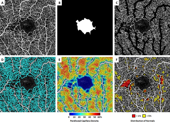
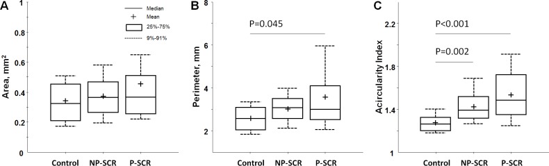
This is a multi-institutional retrospective study of patients with various stages of SCR compared with healthy controls. Parafoveal OCT-A images obtained using a commercial spectral domain-OCT system were reviewed. Foveal-centred 3×3 mm full vascular slab OCT-As were used for image processing and data analysis. FAZ area, perimeter, and acircularity index were determined on the OCT-A image after manual delineation of the FAZ border. Quadrant-based parafoveal capillary density and per cent area deviating from normal distribution were also measured.

RESULTS

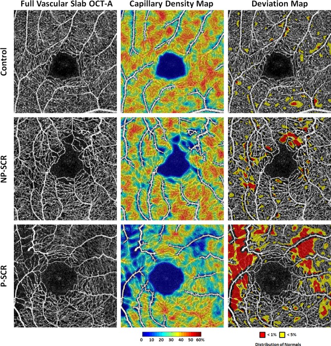
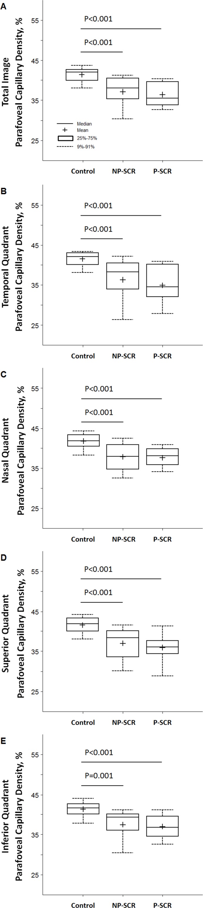
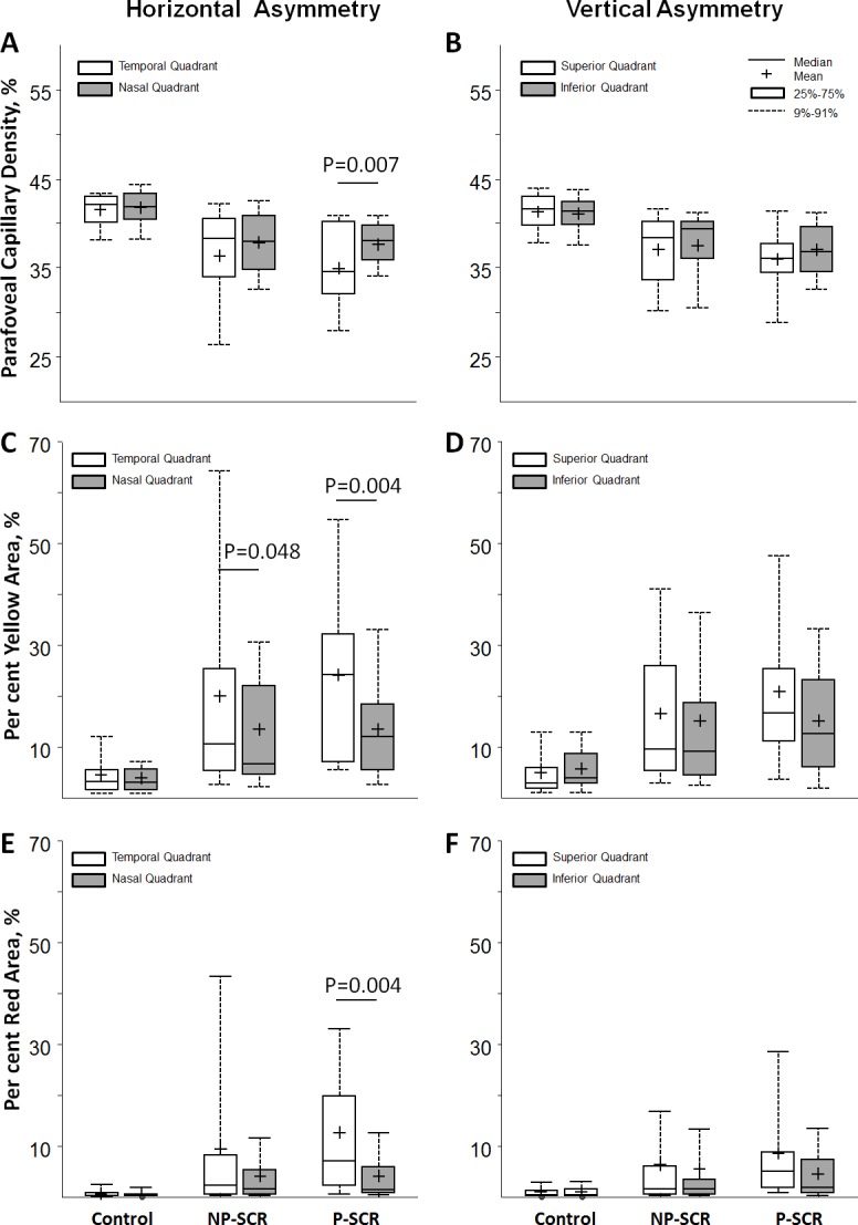
Fifty-two patients with SCR (33 non-proliferative and 19 proliferative) and 20 age and race-matched healthy controls were included. One randomly selected eye per study participant was analysed. FAZ perimeter and acircularity index were significantly greater in SCR eyes when compared with the controls. While parafoveal capillary density was significantly lower, per cent area deviated from normal distribution was significantly higher in SCR eyes than that of the control. However, no statistically significant difference between the two SCR stages was observed. In quadrant-based analysis, the temporal quadrant showed greater parafoveal capillary dropout due to SCR, with the most profound effect in patients with proliferative SCR.

CONCLUSIONS

Abnormal FAZ morphology and altered parafoveal capillary perfusion were found in patients with SCR. Our customised OCT-A image analysis method uniquely highlights significant quantitative alterations in perfusion density mapping in a qualitative display, with minimal obscuration of OCT-A image detail.

摘要

背景/目的:利用光相干断层扫描血管造影术(OCT-A)评估各种镰状细胞视网膜病变(SCR)患者的中心凹无血管区(FAZ)形态和旁中心凹毛细血管灌注。

方法

这是一项多中心回顾性研究,比较了各种阶段 SCR 患者与健康对照者。使用商业谱域-OCT 系统获得旁中心凹 OCT-A 图像。使用 3×3mm 全血管 OCT-A 进行图像处理和数据分析。在手动描绘 FAZ 边界后,在 OCT-A 图像上确定 FAZ 区域、周长和非圆度指数。还测量了基于象限的旁中心凹毛细血管密度和偏离正常分布的百分比区域。

结果

共纳入 52 例 SCR 患者(33 例非增殖性和 19 例增殖性)和 20 例年龄和种族匹配的健康对照者。每位研究参与者随机选择一只眼进行分析。与对照组相比,SCR 眼的 FAZ 周长和非圆度指数明显更大。而旁中心凹毛细血管密度明显降低,SCR 眼偏离正常分布的百分比区域明显高于对照组。然而,两个 SCR 阶段之间没有统计学上的显著差异。在基于象限的分析中,由于 SCR,颞象限的旁中心凹毛细血管丢失更明显,而增殖性 SCR 患者的影响最为严重。

结论

在 SCR 患者中发现了异常的 FAZ 形态和旁中心凹毛细血管灌注改变。我们的定制化 OCT-A 图像分析方法独特地突出了在定性显示中灌注密度图的显著定量改变,同时最小化了 OCT-A 图像细节的遮挡。

https://cdn.ncbi.nlm.nih.gov/pmc/blobs/2d39/7147182/a014cbdc2d8a/bjophthalmol-2019-314567f05.jpg
https://cdn.ncbi.nlm.nih.gov/pmc/blobs/2d39/7147182/23f2b2b5fb59/bjophthalmol-2019-314567f01.jpg
https://cdn.ncbi.nlm.nih.gov/pmc/blobs/2d39/7147182/beabb282603b/bjophthalmol-2019-314567f02.jpg
https://cdn.ncbi.nlm.nih.gov/pmc/blobs/2d39/7147182/b5a18e98b856/bjophthalmol-2019-314567f03.jpg
https://cdn.ncbi.nlm.nih.gov/pmc/blobs/2d39/7147182/6204feae04a2/bjophthalmol-2019-314567f04.jpg
https://cdn.ncbi.nlm.nih.gov/pmc/blobs/2d39/7147182/a014cbdc2d8a/bjophthalmol-2019-314567f05.jpg

相似文献

1
Foveal avascular zone morphology and parafoveal capillary perfusion in sickle cell retinopathy.镰状细胞视网膜病变中中心凹无血管区形态和旁中心凹毛细血管灌注。
Br J Ophthalmol. 2020 Apr;104(4):473-479. doi: 10.1136/bjophthalmol-2019-314567. Epub 2019 Jul 23.
2
Interocular asymmetry of foveal avascular zone morphology and parafoveal capillary density in sickle cell retinopathy.镰状细胞视网膜病变中黄斑中心凹无血管区形态和旁中心凹毛细血管密度的眼间不对称性。
PLoS One. 2020 Jun 10;15(6):e0234151. doi: 10.1371/journal.pone.0234151. eCollection 2020.
3
CORRELATION OF MICROVASCULAR STRUCTURES ON OPTICAL COHERENCE TOMOGRAPHY ANGIOGRAPHY WITH VISUAL ACUITY IN RETINAL VEIN OCCLUSION.视网膜静脉阻塞患者光学相干断层扫描血管造影微血管结构与视力的相关性
Retina. 2017 Sep;37(9):1700-1709. doi: 10.1097/IAE.0000000000001403.
4
[Correlation of capillary plexus with visual acuity in idiopathic macular epiretinal membrane eyes using optical coherence tomography angiography].[利用光学相干断层扫描血管造影术研究特发性黄斑视网膜前膜眼中毛细血管丛与视力的相关性]
Zhonghua Yan Ke Za Zhi. 2019 Oct 11;55(10):757-762. doi: 10.3760/cma.j.issn.0412-4081.2019.10.006.
5
Optical coherence tomography angiography analysis of foveal microvascular changes and inner retinal layer thinning in patients with diabetes.糖尿病患者黄斑区微血管改变及内层视网膜变薄的光相干断层扫描血管造影分析。
Br J Ophthalmol. 2018 Sep;102(9):1226-1231. doi: 10.1136/bjophthalmol-2017-311149. Epub 2017 Dec 19.
6
Parafoveal Microvascular Alterations in Ocular and Non-Ocular Behҫet's Disease Evaluated With Optical Coherence Tomography Angiography.用光学相干断层血管造影术评估眼部和非眼部贝赫切特病的旁中心区微血管改变。
Invest Ophthalmol Vis Sci. 2021 Mar 1;62(3):8. doi: 10.1167/iovs.62.3.8.
7
Quantitative optical coherence tomography angiography of macular vascular structure and foveal avascular zone in glaucoma.青光眼黄斑血管结构及中心凹无血管区的定量光学相干断层扫描血管造影
PLoS One. 2017 Sep 21;12(9):e0184948. doi: 10.1371/journal.pone.0184948. eCollection 2017.
8
Imaging Foveal Microvasculature: Optical Coherence Tomography Angiography Versus Adaptive Optics Scanning Light Ophthalmoscope Fluorescein Angiography.黄斑区微血管成像:光学相干断层扫描血管造影术与自适应光学扫描激光眼底镜荧光血管造影术的对比
Invest Ophthalmol Vis Sci. 2016 Jul 1;57(9):OCT130-40. doi: 10.1167/iovs.15-18932.
9
VARIABILITY IN FOVEAL AVASCULAR ZONE AND CAPILLARY DENSITY USING OPTICAL COHERENCE TOMOGRAPHY ANGIOGRAPHY MACHINES IN HEALTHY EYES.使用光学相干断层扫描血管造影机检测健康眼睛的黄斑中心凹血管区和毛细血管密度的变异性
Retina. 2017 Nov;37(11):2102-2111. doi: 10.1097/IAE.0000000000001458.
10
Foveal ganglion cell layer damage in ischemic diabetic maculopathy: correlation of optical coherence tomographic and anatomic changes.缺血性糖尿病性黄斑病变中的黄斑神经节细胞层损伤:光学相干断层扫描与解剖学变化的相关性
Ophthalmology. 2009 Oct;116(10):1949-59.e8. doi: 10.1016/j.ophtha.2009.06.066. Epub 2009 Aug 21.

引用本文的文献

1
Optical coherence tomography angiography as a tool for diagnosis and monitoring of sickle cell related eye disease: a systematic review and meta-analysis.光学相干断层扫描血管造影术作为镰状细胞相关眼病诊断和监测工具的系统评价与荟萃分析
Eye (Lond). 2025 May 22. doi: 10.1038/s41433-025-03814-1.
2
Preferential Sites of Retinal Capillary Occlusion in Sickle Cell Disease.镰状细胞病视网膜毛细血管阻塞的好发部位
Invest Ophthalmol Vis Sci. 2025 Jan 2;66(1):57. doi: 10.1167/iovs.66.1.57.
3
MACULAR NEURODEGENERATIVE AND VASCULAR CHANGES ON OPTICAL COHERENCE TOMOGRAPHIC ANGIOGRAPHY IN SICKLE CELL DISEASE ARE NOT RELATED TO ITS OCULAR AND SYSTEMIC COMPLICATIONS.

本文引用的文献

1
Novel Development of Parafoveal Capillary Density Deviation Mapping using an Age-Group and Eccentricity Matched Normative OCT Angiography Database.利用年龄组和偏心度匹配的规范光学相干断层扫描血管造影数据库进行旁中心凹毛细血管密度偏差映射的新进展。
Transl Vis Sci Technol. 2019 May 1;8(3):1. doi: 10.1167/tvst.8.3.1. eCollection 2019 May.
2
Correlation of Ultra-Widefield Fluorescein Angiography and OCT Angiography in Sickle Cell Retinopathy.镰状细胞视网膜病变中超宽视野荧光素血管造影与光学相干断层扫描血管造影的相关性
Ophthalmol Retina. 2018 Jun;2(6):599-605. doi: 10.1016/j.oret.2017.10.011. Epub 2017 Dec 9.
3
Variability of Foveal Avascular Zone Metrics Derived From Optical Coherence Tomography Angiography Images.
镰状细胞病患者光学相干断层扫描血管造影中的黄斑神经退行性和血管变化与其眼部及全身并发症无关。
Retina. 2024 Dec 1;44(12):2185-2195. doi: 10.1097/IAE.0000000000004247.
4
Optical coherence tomography angiography of the macula of high myopia in children and adolescents.儿童及青少年高度近视黄斑区的光学相干断层扫描血管造影
Int J Retina Vitreous. 2024 Feb 5;10(1):17. doi: 10.1186/s40942-024-00532-w.
5
Peak Cone Density Predicted from Outer Segment Length Measured on Optical Coherence Tomography.基于光学相干断层扫描测量的外节段长度预测的峰锥密度。
Curr Eye Res. 2024 Mar;49(3):314-324. doi: 10.1080/02713683.2023.2289853. Epub 2023 Dec 26.
6
Insights into Sickle Cell Disease through the Retinal Microvasculature: Adaptive Optics Scanning Light Ophthalmoscopy Correlates of Clinical OCT Angiography.通过视网膜微血管系统洞察镰状细胞病:自适应光学扫描激光检眼镜与临床光学相干断层扫描血管造影的相关性
Ophthalmol Sci. 2022 Jul 12;2(4):100196. doi: 10.1016/j.xops.2022.100196. eCollection 2022 Dec.
7
Optical coherence tomography angiography for the characterisation of retinal microvasculature alterations in pregnant patients with anaemia: a nested case‒control study.光学相干断层扫描血管造影术用于贫血孕妇视网膜微血管改变的特征分析:巢式病例对照研究。
Br J Ophthalmol. 2023 Dec 18;108(1):117-123. doi: 10.1136/bjo-2022-321781.
8
Quantification of intermittent retinal capillary perfusion in sickle cell disease.镰状细胞病中间歇性视网膜毛细血管灌注的定量分析
Biomed Opt Express. 2021 Apr 21;12(5):2825-2840. doi: 10.1364/BOE.418874. eCollection 2021 May 1.
9
Artificial intelligence for improving sickle cell retinopathy diagnosis and management.人工智能在镰状细胞性视网膜病变诊断和管理中的应用。
Eye (Lond). 2021 Oct;35(10):2675-2684. doi: 10.1038/s41433-021-01556-4. Epub 2021 May 6.
10
Analysis of the foveal microvasculature in sickle cell disease using swept-source optical coherence tomography angiography.应用扫频源光相干断层扫描血管造影分析镰状细胞病的中心凹微血管。
Sci Rep. 2020 Jul 16;10(1):11795. doi: 10.1038/s41598-020-68625-8.
基于光学相干断层扫描血管造影图像得出的黄斑无血管区测量指标的变异性
Transl Vis Sci Technol. 2018 Oct 1;7(5):20. doi: 10.1167/tvst.7.5.20. eCollection 2018 Sep.
4
A method for age-matched OCT angiography deviation mapping in the assessment of disease- related changes to the radial peripapillary capillaries.一种用于年龄匹配 OCT 血管造影偏差映射的方法,用于评估与放射状视盘毛细血管相关的疾病变化。
PLoS One. 2018 May 24;13(5):e0197062. doi: 10.1371/journal.pone.0197062. eCollection 2018.
5
Quantitative characteristics of sickle cell retinopathy in optical coherence tomography angiography.光学相干断层扫描血管造影中镰状细胞视网膜病变的定量特征
Biomed Opt Express. 2017 Feb 23;8(3):1741-1753. doi: 10.1364/BOE.8.001741. eCollection 2017 Mar 1.
6
Axial Length Variation Impacts on Superficial Retinal Vessel Density and Foveal Avascular Zone Area Measurements Using Optical Coherence Tomography Angiography.眼轴长度变化对使用光学相干断层扫描血管造影术测量视网膜浅表血管密度和黄斑无血管区面积的影响。
Invest Ophthalmol Vis Sci. 2017 Jun 1;58(7):3065-3072. doi: 10.1167/iovs.17-21551.
7
Assessing the Accuracy of Foveal Avascular Zone Measurements Using Optical Coherence Tomography Angiography: Segmentation and Scaling.使用光学相干断层扫描血管造影术评估黄斑无血管区测量的准确性:分割与缩放
Transl Vis Sci Technol. 2017 Jun 9;6(3):16. doi: 10.1167/tvst.6.3.16. eCollection 2017 Jun.
8
Acircularity index and axis ratio of the foveal avascular zone in diabetic eyes and healthy controls measured by optical coherence tomography angiography.通过光学相干断层扫描血管造影测量糖尿病患者眼睛和健康对照者的黄斑无血管区的圆度指数和轴比。
Vision Res. 2017 Oct;139:177-186. doi: 10.1016/j.visres.2016.09.019. Epub 2017 Feb 27.
9
Evaluation of Macular Vascular Abnormalities Identified by Optical Coherence Tomography Angiography in Sickle Cell Disease.镰状细胞病中光学相干断层扫描血管造影术所识别的黄斑血管异常的评估
Am J Ophthalmol. 2017 May;177:90-99. doi: 10.1016/j.ajo.2017.02.007. Epub 2017 Feb 14.
10
VARIABILITY IN FOVEAL AVASCULAR ZONE AND CAPILLARY DENSITY USING OPTICAL COHERENCE TOMOGRAPHY ANGIOGRAPHY MACHINES IN HEALTHY EYES.使用光学相干断层扫描血管造影机检测健康眼睛的黄斑中心凹血管区和毛细血管密度的变异性
Retina. 2017 Nov;37(11):2102-2111. doi: 10.1097/IAE.0000000000001458.