Rotterdam Ophthalmic Institute, Rotterdam, The Netherlands.
Uveitis Service, Rotterdam Eye Hospital, Rotterdam, The Netherlands.
Invest Ophthalmol Vis Sci. 2021 Mar 1;62(3):8. doi: 10.1167/iovs.62.3.8.
To compare quantitative optical coherence tomography angiography (OCT-A) measurements of the parafoveal microvasculature in retinal capillary plexuses among Behҫet uveitis (BU) patients, non-ocular Behҫet's disease (NOBD) patients, and healthy volunteers (HVs).
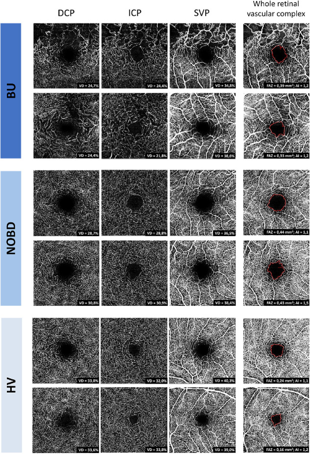
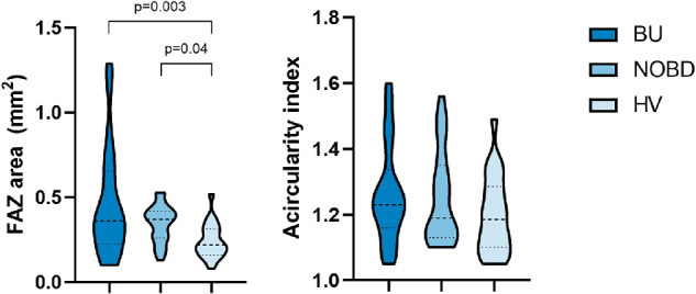
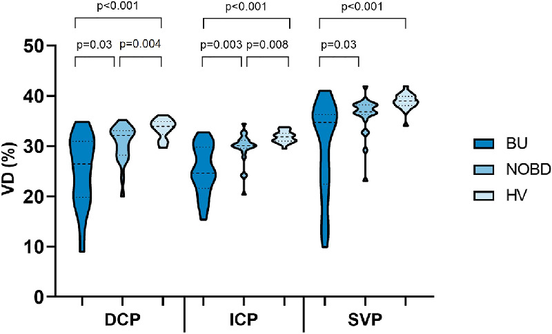
Sixty-eight subjects were enrolled in this prospective observational cross-sectional study. OCT-A imaging was performed using the Heidelberg Engineering Spectralis OCT. A custom algorithm was developed to calculate the vessel density (VD) in three retinal vascular layers: deep capillary plexus, intermediate capillary plexus, and superficial vascular plexus. The foveal avascular zone (FAZ) and acircularity index were calculated for the whole retinal vascular complex.
We analyzed one eye from 21 BU patients (age, 51 ± 10 years), 23 NOBD patients (age, 48 ± 14 years), and 22 HVs (age, 44 ± 13 years). One-way multivariate analysis of covariance showed a statistically significant difference in VD among the three groups when combining the layers after controlling for scan quality (P < 0.001). The VD was lowest in the BU group and highest in the HV group in all layers. The FAZ area was also statistically significant different among the groups (P < 0.005), with the largest FAZ areas in BU patients and smallest FAZ areas in the HV group. However, no statistically significant difference was found for the acircularity index.
The parafoveal microvasculature is affected not only in BU patients but also in NOBD patients. Most deviations in the retinal microcirculation in Behҫet patients were found in the deeper layers of the retina by using the quantitative VD measurement.
比较 Behçet 葡萄膜炎(BU)患者、非眼部 Behçet 病(NOBD)患者和健康志愿者(HV)视网膜毛细血管丛旁黄斑区微血管的定量光相干断层扫描血管造影(OCT-A)测量值。
本前瞻性观察性横断面研究纳入了 68 名受试者。使用海德堡工程 Spectralis OCT 进行 OCT-A 成像。开发了一种自定义算法来计算三个视网膜血管层的血管密度(VD):深层毛细血管丛、中间毛细血管丛和浅层血管丛。计算整个视网膜血管复合体的黄斑无血管区(FAZ)和非圆度指数。
我们分析了 21 名 BU 患者(年龄 51 ± 10 岁)、23 名 NOBD 患者(年龄 48 ± 14 岁)和 22 名 HV(年龄 44 ± 13 岁)的一只眼。在控制扫描质量后,对所有层进行单因素多变量协方差分析,结果显示三组间 VD 存在统计学差异(P < 0.001)。结合各层后,VD 在 BU 组中最低,在 HV 组中最高。各组间 FAZ 面积也存在统计学差异(P < 0.005),BU 患者的 FAZ 面积最大,HV 组的 FAZ 面积最小。然而,对于非圆度指数,未发现统计学差异。
旁黄斑区微血管不仅受 BU 患者影响,也受 NOBD 患者影响。使用定量 VD 测量发现,Behçet 患者视网膜微循环的大多数异常发生在视网膜的深层。