• 文献检索
  • 文档翻译
  • 深度研究
  • 学术资讯
  • Suppr Zotero 插件Zotero 插件
  • 邀请有礼
  • 套餐&价格
  • 历史记录
应用&插件
Suppr Zotero 插件Zotero 插件浏览器插件Mac 客户端Windows 客户端微信小程序
定价
高级版会员购买积分包购买API积分包
服务
文献检索文档翻译深度研究API 文档MCP 服务
关于我们
关于 Suppr公司介绍联系我们用户协议隐私条款
关注我们

Suppr 超能文献

核心技术专利:CN118964589B侵权必究
粤ICP备2023148730 号-1Suppr @ 2026

文献检索

告别复杂PubMed语法,用中文像聊天一样搜索,搜遍4000万医学文献。AI智能推荐,让科研检索更轻松。

立即免费搜索

文件翻译

保留排版,准确专业,支持PDF/Word/PPT等文件格式,支持 12+语言互译。

免费翻译文档

深度研究

AI帮你快速写综述,25分钟生成高质量综述,智能提取关键信息,辅助科研写作。

立即免费体验

一种用于年龄匹配 OCT 血管造影偏差映射的方法,用于评估与放射状视盘毛细血管相关的疾病变化。

A method for age-matched OCT angiography deviation mapping in the assessment of disease- related changes to the radial peripapillary capillaries.

机构信息

Department of Ophthalmology, New York Eye and Ear Infirmary of Mount Sinai, New York, NY, United States of America.

Department of Ophthalmology, State University of New York Downstate Medical Center, Brooklyn, NY, United States of America.

出版信息

PLoS One. 2018 May 24;13(5):e0197062. doi: 10.1371/journal.pone.0197062. eCollection 2018.

DOI:10.1371/journal.pone.0197062
PMID:29795576
原文链接:https://pmc.ncbi.nlm.nih.gov/articles/PMC5993123/
Abstract

PURPOSE

To present a method for age-matched deviation mapping in the assessment of disease-related changes to the radial peripapillary capillaries (RPCs).

METHODS

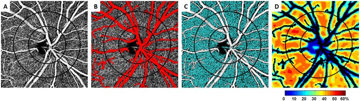
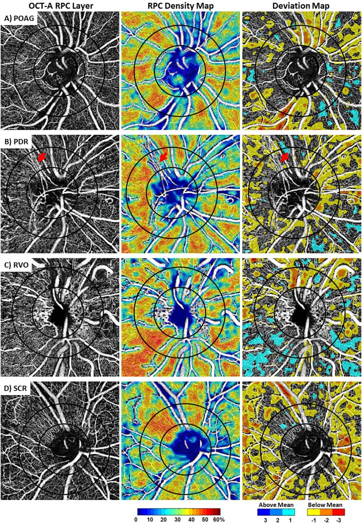
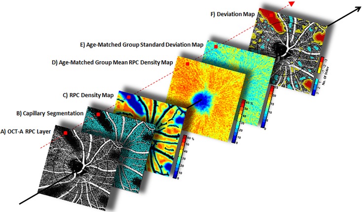
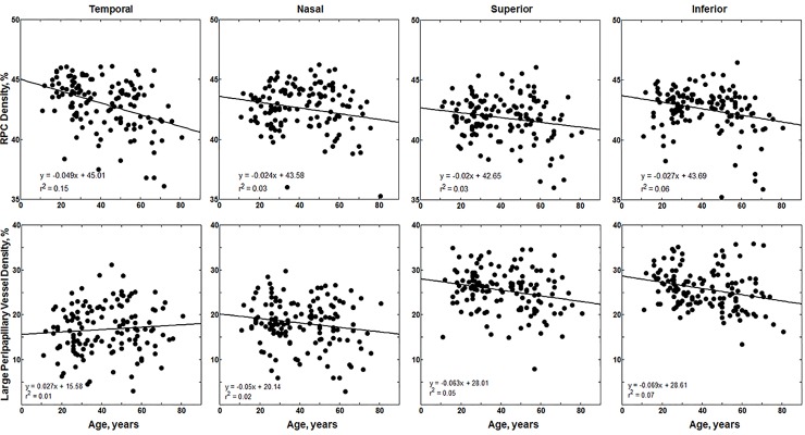
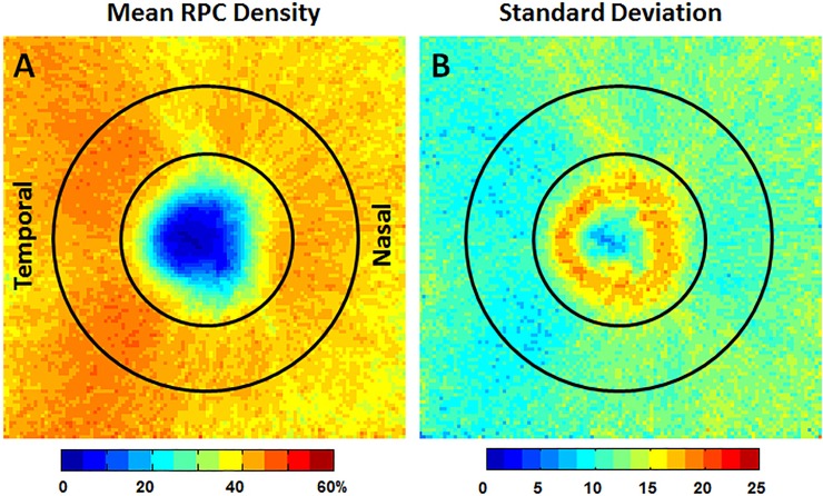
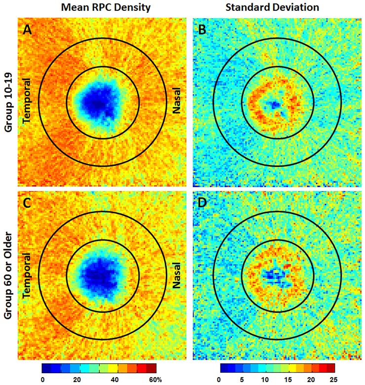
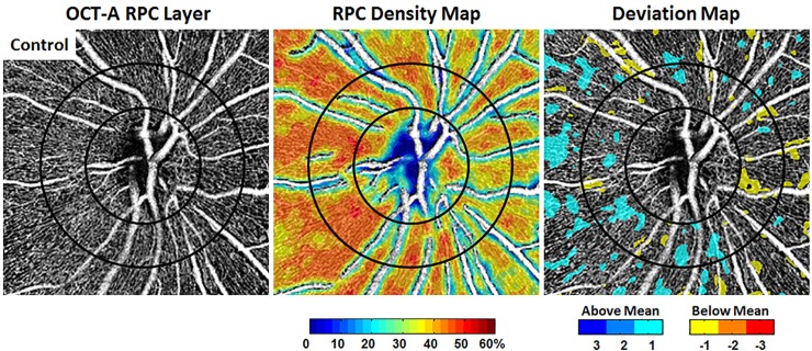
We reviewed 4.5x4.5mm en face peripapillary OCT-A scans of 133 healthy control eyes (133 subjects, mean 41.5 yrs, range 11-82 yrs) and 4 eyes with distinct retinal pathologies, obtained using spectral-domain optical coherence tomography angiography. Statistical analysis was performed to evaluate the impact of age on RPC perfusion densities. RPC density group mean and standard deviation maps were generated for each decade of life. Deviation maps were created for the diseased eyes based on these maps. Large peripapillary vessel (LPV; noncapillary vessel) perfusion density was also studied for impact of age.

RESULTS

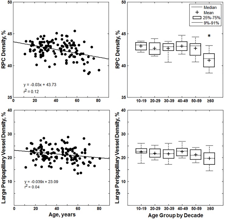
Average healthy RPC density was 42.5±1.47%. ANOVA and pairwise Tukey-Kramer tests showed that RPC density in the ≥60yr group was significantly lower compared to RPC density in all younger decades of life (p<0.01). Average healthy LPV density was 21.5±3.07%. Linear regression models indicated that LPV density decreased with age, however ANOVA and pairwise Tukey-Kramer tests did not reach statistical significance. Deviation mapping enabled us to quantitatively and visually elucidate the significance of RPC density changes in disease.

CONCLUSIONS

It is important to consider changes that occur with aging when analyzing RPC and LPV density changes in disease. RPC density, coupled with age-matched deviation mapping techniques, represents a potentially clinically useful method in detecting changes to peripapillary perfusion in disease.

摘要

目的

提出一种用于评估与疾病相关的视盘周围毛细血管(RPC)变化的年龄匹配偏差映射方法。

方法

我们回顾了 133 只健康对照眼(133 例,平均年龄 41.5 岁,范围 11-82 岁)和 4 只具有明显视网膜病变的眼的 4.5x4.5mm 额面视盘 OCT-A 扫描。使用谱域光相干断层扫描血管造影术获得了统计学分析,以评估年龄对 RPC 灌注密度的影响。为每个生命十年生成 RPC 密度组平均值和标准偏差图。根据这些图为患病眼创建偏差图。还研究了大视盘血管(LPV;非毛细血管血管)灌注密度对年龄的影响。

结果

健康 RPC 平均密度为 42.5±1.47%。方差分析和两两 Tukey-Kramer 检验表明,≥60 岁组的 RPC 密度明显低于所有年轻年龄段的 RPC 密度(p<0.01)。健康 LPV 平均密度为 21.5±3.07%。线性回归模型表明 LPV 密度随年龄下降,但方差分析和两两 Tukey-Kramer 检验未达到统计学意义。偏差映射使我们能够定量和直观地阐明疾病中 RPC 密度变化的意义。

结论

在分析疾病中 RPC 和 LPV 密度变化时,考虑与衰老相关的变化非常重要。RPC 密度结合年龄匹配偏差映射技术,代表了一种在疾病中检测视盘周围灌注变化的潜在临床有用方法。

https://cdn.ncbi.nlm.nih.gov/pmc/blobs/c531/5993123/25a429d536f8/pone.0197062.g008.jpg
https://cdn.ncbi.nlm.nih.gov/pmc/blobs/c531/5993123/23c6163d9c9e/pone.0197062.g001.jpg
https://cdn.ncbi.nlm.nih.gov/pmc/blobs/c531/5993123/16794492452c/pone.0197062.g002.jpg
https://cdn.ncbi.nlm.nih.gov/pmc/blobs/c531/5993123/63392fdc5555/pone.0197062.g003.jpg
https://cdn.ncbi.nlm.nih.gov/pmc/blobs/c531/5993123/1e9129b735e8/pone.0197062.g004.jpg
https://cdn.ncbi.nlm.nih.gov/pmc/blobs/c531/5993123/0196dfa76b46/pone.0197062.g005.jpg
https://cdn.ncbi.nlm.nih.gov/pmc/blobs/c531/5993123/b1137d3a38c9/pone.0197062.g006.jpg
https://cdn.ncbi.nlm.nih.gov/pmc/blobs/c531/5993123/f1e926c351d4/pone.0197062.g007.jpg
https://cdn.ncbi.nlm.nih.gov/pmc/blobs/c531/5993123/25a429d536f8/pone.0197062.g008.jpg

相似文献

1
A method for age-matched OCT angiography deviation mapping in the assessment of disease- related changes to the radial peripapillary capillaries.一种用于年龄匹配 OCT 血管造影偏差映射的方法,用于评估与放射状视盘毛细血管相关的疾病变化。
PLoS One. 2018 May 24;13(5):e0197062. doi: 10.1371/journal.pone.0197062. eCollection 2018.
2
Effect of Scan Size on Glaucoma Diagnostic Performance Using OCT Angiography En Face Images of the Radial Peripapillary Capillaries.基于 OCT 血管成像的放射状视神经周围毛细血管的扫描大小对青光眼诊断性能的影响。
J Glaucoma. 2019 May;28(5):465-472. doi: 10.1097/IJG.0000000000001216.
3
Assessment of Perfused Peripapillary Capillaries and Peripapillary Capillary Density Maps in Glaucoma Patients.青光眼患者的灌注神经纤维层毛细血管和神经纤维层毛细血管密度图评估。
Med Arch. 2020 Aug;74(4):275-278. doi: 10.5455/medarh.2020.74.275-278.
4
Quantitative Optical Coherence Tomography Angiography of Radial Peripapillary Capillaries in Glaucoma, Glaucoma Suspect, and Normal Eyes.青光眼、疑似青光眼和正常眼的视盘周围放射状毛细血管的定量光学相干断层扫描血管造影
Am J Ophthalmol. 2016 Oct;170:41-49. doi: 10.1016/j.ajo.2016.07.015. Epub 2016 Jul 25.
5
Quantitative Analysis of Microvasculature in Macular and Peripapillary Regions in Early Primary Open-Angle Glaucoma.早期原发性开角型青光眼黄斑及视盘周围区域微血管定量分析。
Curr Eye Res. 2020 May;45(5):629-635. doi: 10.1080/02713683.2019.1676912. Epub 2019 Oct 14.
6
Correlation of optic disc morphometry and optic disc microvasculature assessed with optical coherence tomography angiography.利用光学相干断层扫描血管造影评估视盘形态测量和视盘微血管。
Can J Ophthalmol. 2018 Dec;53(6):595-599. doi: 10.1016/j.jcjo.2018.01.026. Epub 2018 Mar 9.
7
A comparison of two optical coherence tomography-angiography devices in pseudoexfoliation glaucoma versus primary open-angle glaucoma and healthy subjects.两种光学相干断层扫描血管造影设备在假性剥脱性青光眼与原发性开角型青光眼及健康受试者中的比较。
Eur J Ophthalmol. 2019 Nov;29(6):636-644. doi: 10.1177/1120672118805882. Epub 2018 Oct 14.
8
Quantitative Analysis of the RPC Vessel Density and the RNFL Thickness in Patients with Type 2 Diabetes Mellitus by Using OCT Angiography.应用光学相干断层血管成像术定量分析 2 型糖尿病患者视网膜毛细血管密度和神经纤维层厚度。
Ophthalmic Res. 2021;64(6):951-959. doi: 10.1159/000517145. Epub 2021 Jul 20.
9
Immediate Changes in Peripapillary Retinal Vasculature after Intraocular Pressure Elevation -an Optical Coherence Tomography Angiography Study.眼压升高后视乳头周围视网膜血管的即时变化——一项光相干断层扫描血管造影研究。
Curr Eye Res. 2020 Jun;45(6):749-756. doi: 10.1080/02713683.2019.1695843. Epub 2019 Nov 27.
10
Correlation of retinal sensitivity in microperimetry with vascular density in optical coherence tomography angiography in primary open-angle glaucoma.原发性开角型青光眼微视野检查中视网膜敏感度与光学相干断层扫描血管造影中血管密度的相关性
PLoS One. 2020 Jul 6;15(7):e0235571. doi: 10.1371/journal.pone.0235571. eCollection 2020.

引用本文的文献

1
Aging affects retinal oxygen and blood flow metrics in mice.衰老会影响小鼠视网膜的氧气和血流指标。
Exp Eye Res. 2025 Jun;255:110363. doi: 10.1016/j.exer.2025.110363. Epub 2025 Mar 26.
2
Vascular density normative data of radial peripapillary capillary plexus in healthy Caucasian subjects.健康高加索受试者视乳头周围视网膜毛细血管丛的血管密度标准数据。
Graefes Arch Clin Exp Ophthalmol. 2025 Jan;263(1):97-103. doi: 10.1007/s00417-024-06623-6. Epub 2024 Aug 30.
3
Assessing the Sensitivity of OCT-A Retinal Vasculature Metrics.评估 OCT-A 视网膜血管测量指标的敏感性。

本文引用的文献

1
Age-Related Alterations in the Retinal Microvasculature, Microcirculation, and Microstructure.视网膜微血管系统、微循环和微结构的年龄相关性改变。
Invest Ophthalmol Vis Sci. 2017 Jul 1;58(9):3804-3817. doi: 10.1167/iovs.17-21460.
2
Quantitative OCT Angiography of the Retinal Microvasculature and the Choriocapillaris in Myopic Eyes.近视眼中视网膜微血管和脉络膜毛细血管的定量光学相干断层扫描血管造影
Invest Ophthalmol Vis Sci. 2017 Apr 1;58(4):2063-2069. doi: 10.1167/iovs.16-21289.
3
Peripapillary Retinal Nerve Fiber Layer Vascular Microcirculation in Eyes With Glaucoma and Single-Hemifield Visual Field Loss.
Transl Vis Sci Technol. 2023 Aug 1;12(8):2. doi: 10.1167/tvst.12.8.2.
4
Age-related assessment of foveal avascular zone and surrounding capillary networks with swept source optical coherence tomography angiography in healthy eyes.年龄相关性黄斑中心凹无血管区及其周围毛细血管网的扫频源光相干断层扫描血管造影评估。
Eye (Lond). 2022 Oct;36(10):1857-1864. doi: 10.1038/s41433-022-02146-8. Epub 2022 Aug 11.
5
Thyroid-Associated Peripapillary Vascular Remodelling - A Novel Area for Research? [Letter].甲状腺相关性视乳头周围血管重塑——一个新的研究领域?[信函]
Clin Ophthalmol. 2022 May 12;16:1475-1476. doi: 10.2147/OPTH.S372789. eCollection 2022.
6
A Pilot Study of Subclinical Non-Capillary Peripapillary Perfusion Changes in Thyroid-Related Orbitopathy Detected Using Optical Coherence Tomography Angiography.一项利用光学相干断层扫描血管造影术检测甲状腺相关性眼病亚临床非毛细血管视乳头周围灌注变化的初步研究。
Clin Ophthalmol. 2022 Mar 20;16:867-875. doi: 10.2147/OPTH.S356631. eCollection 2022.
7
Combined wide-field optical coherence tomography angiography density map for high myopic glaucoma detection.用于高度近视性青光眼检测的宽视野光学相干断层扫描血管造影密度图的联合应用。
Sci Rep. 2021 Nov 11;11(1):22034. doi: 10.1038/s41598-021-01661-0.
8
Retinal alterations in patients with Lafora disease.拉福拉病患者的视网膜改变。
Am J Ophthalmol Case Rep. 2021 Jun 15;23:101146. doi: 10.1016/j.ajoc.2021.101146. eCollection 2021 Sep.
9
Optic disc and peripapillary vessel density measured with optical coherence tomography angiography and its associations in Chinese adults: a large population-based study.基于光学相干断层扫描血管造影术测量的中国成年人视盘和视乳头周围血管密度及其相关性:一项基于大人群的研究
Br J Ophthalmol. 2022 Oct;106(10):1411-1416. doi: 10.1136/bjophthalmol-2020-318199. Epub 2021 May 20.
10
Interocular asymmetry of foveal avascular zone morphology and parafoveal capillary density in sickle cell retinopathy.镰状细胞视网膜病变中黄斑中心凹无血管区形态和旁中心凹毛细血管密度的眼间不对称性。
PLoS One. 2020 Jun 10;15(6):e0234151. doi: 10.1371/journal.pone.0234151. eCollection 2020.
青光眼伴单眼半视野缺损患者的视乳头周围视网膜神经纤维层血管微循环
JAMA Ophthalmol. 2017 May 1;135(5):461-468. doi: 10.1001/jamaophthalmol.2017.0261.
4
Optical Coherence Angiographic Demonstration of Retinal Changes From Chronic Optic Neuropathies.慢性视神经病变视网膜变化的光学相干血管造影显示
Neuroophthalmology. 2017 Jan 27;41(2):76-83. doi: 10.1080/01658107.2016.1275703. eCollection 2017 Apr.
5
Optical Coherence Tomography Angiography in Nonarteritic Anterior Ischemic Optic Neuropathy.光学相干断层扫描血管造影术在非动脉炎性前部缺血性视神经病变中的应用
J Neuroophthalmol. 2017 Dec;37(4):358-364. doi: 10.1097/WNO.0000000000000493.
6
Radial Peripapillary Capillary Density Measurement Using Optical Coherence Tomography Angiography in Early Glaucoma.在早期青光眼患者中使用光学相干断层扫描血管造影术测量视乳头周围视网膜毛细血管密度
J Glaucoma. 2017 May;26(5):438-443. doi: 10.1097/IJG.0000000000000649.
7
Peripapillary and Macular Vessel Density in Patients with Glaucoma and Single-Hemifield Visual Field Defect.青光眼和单半视野视觉缺损患者的视乳头周围和黄斑区血管密度
Ophthalmology. 2017 May;124(5):709-719. doi: 10.1016/j.ophtha.2017.01.004. Epub 2017 Feb 10.
8
Detailed Vascular Anatomy of the Human Retina by Projection-Resolved Optical Coherence Tomography Angiography.基于层析分辨光学相干断层血管造影术的人视网膜详细血管解剖。
Sci Rep. 2017 Feb 10;7:42201. doi: 10.1038/srep42201.
9
Peripapillary perfused capillary density in primary openangle glaucoma across disease stage an optical coherence tomography angiography study.原发性开角型青光眼不同疾病阶段的视乳头周围灌注毛细血管密度:一项光学相干断层扫描血管造影研究
Br J Ophthalmol. 2017 Sep;101(9):1261-1268. doi: 10.1136/bjophthalmol-2016-309642. Epub 2017 Feb 1.
10
OCT angiography of acute non-arteritic anterior ischemic optic neuropathy.急性非动脉炎性前部缺血性视神经病变的光学相干断层扫描血管造影
J Fr Ophtalmol. 2017 Feb;40(2):102-109. doi: 10.1016/j.jfo.2016.09.020. Epub 2017 Jan 20.