Department of Global Health, Faculty of Medicine and Health Sciences, African Cancer Institute, Stellenbosch University, Cape Town, 8000, South Africa.
Department of Physiological Sciences, Faculty of Science, Stellenbosch University, Stellenbosch, 7600, South Africa.
BMC Cancer. 2019 Aug 1;19(1):757. doi: 10.1186/s12885-019-5939-z.
Doxorubicin is currently the most effective chemotherapeutic drug used to treat breast cancer. It has, however, been shown that doxorubicin can induce drug resistance resulting in poor patient prognosis and survival. Studies reported that the interaction between signalling pathways can promote drug resistance through the induction of proliferation, cell cycle progression and prevention of apoptosis. The aim of this study was therefore to determine the effects of doxorubicin on apoptosis signalling, autophagy, the mitogen-activated protein kinase (MAPK)- and phosphoinositide 3-kinase (PI3K)/Akt signalling pathway, cell cycle control, and regulators of the epithelial-mesenchymal transition (EMT) process in murine breast cancer tumours.
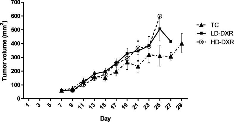
A tumour-bearing mouse model was established by injecting murine E0771 breast cancer cells, suspended in Hank's Balances Salt Solution and Corning® Matrigel® Basement Membrane Matrix, into female C57BL/6 mice. Fourty-seven mice were randomly divided into three groups, namely tumour control (received Hank's Balances Salt Solution), low dose doxorubicin (received total of 6 mg/ml doxorubicin) and high dose doxorubicin (received total of 15 mg/ml doxorubicin) groups. A higher tumour growth rate was, however, observed in doxorubicin-treated mice compared to the untreated controls. We therefore compared the expression levels of markers involved in cell death and survival signalling pathways, by means of western blotting and fluorescence-based immunohistochemistry.
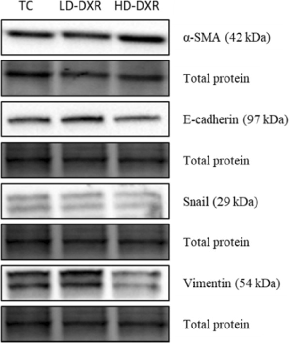
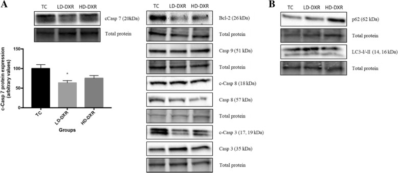
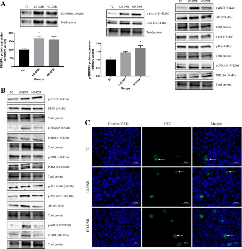
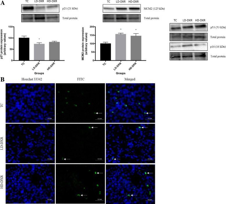
Doxorubicin failed to induce cell death, by means of apoptosis or autophagy, and cell cycle arrest, indicating the occurrence of drug resistance and uncontrolled proliferation. Activation of the MAPK/ extracellular-signal-regulated kinase (ERK) pathway contributed to the resistance observed in treated mice, while no significant changes were found with the PI3K/Akt pathway and other MAPK pathways. Significant changes were also observed in cell cycle p21 and DNA replication minichromosome maintenance 2 proteins. No significant changes in EMT markers were observed after doxorubicin treatment.
Our results suggest that doxorubicin-induced drug resistance and tumour growth can occur through the adaptive role of the MAPK/ERK pathway in an effort to protect tumour cells. Previous studies have shown that the efficacy of doxorubicin can be improved by inhibition of the ERK signalling pathway and thereby treatment failure can be overcome.
多柔比星是目前治疗乳腺癌最有效的化疗药物。然而,已经表明多柔比星可以诱导耐药性,从而导致患者预后和生存不良。研究报告称,信号通路之间的相互作用可以通过促进增殖、细胞周期进展和防止细胞凋亡来促进耐药性。因此,本研究旨在确定多柔比星对凋亡信号、自噬、丝裂原激活蛋白激酶(MAPK)和磷酸肌醇 3-激酶(PI3K)/Akt 信号通路、细胞周期控制以及上皮-间充质转化(EMT)过程调节剂在小鼠乳腺癌肿瘤中的作用。
通过将悬浮在汉克氏平衡盐溶液和康宁®基质胶基底膜基质中的小鼠 E0771 乳腺癌细胞注入雌性 C57BL/6 小鼠中,建立荷瘤小鼠模型。47 只小鼠随机分为三组,即肿瘤对照组(接受汉克氏平衡盐溶液)、低剂量多柔比星组(接受总剂量 6mg/ml 多柔比星)和高剂量多柔比星组(接受总剂量 15mg/ml 多柔比星)。然而,与未治疗的对照组相比,多柔比星治疗的小鼠观察到更高的肿瘤生长速度。因此,我们通过 Western 印迹和荧光免疫组织化学比较了参与细胞死亡和存活信号通路的标志物的表达水平。
多柔比星未能通过细胞凋亡或自噬和细胞周期阻滞诱导细胞死亡,表明发生了耐药性和不受控制的增殖。MAPK/细胞外信号调节激酶(ERK)通路的激活导致了治疗小鼠中观察到的耐药性,而 PI3K/Akt 通路和其他 MAPK 通路没有明显变化。细胞周期 p21 和 DNA 复制微染色体维持 2 蛋白也发生了显著变化。多柔比星治疗后,EMT 标志物无明显变化。
我们的结果表明,多柔比星诱导的耐药性和肿瘤生长可能通过 MAPK/ERK 通路的适应性作用发生,以保护肿瘤细胞。先前的研究表明,通过抑制 ERK 信号通路可以提高多柔比星的疗效,从而克服治疗失败。